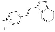
RNA Imaging Probe 1c New
Chemical Name: 3-[(E)-2-(1-Methylpyridin-1-ium-4-yl)ethenyl]indolizine iodide
Purity: ≥98%
Biological Activity
RNA Imaging Probe 1c is a fluorogenic RNA imaging probe. RNA Imaging Probe 1c displays high membrane permeability, strong fluorogenic responses upon binding RNA, compatibility with fluorescence lifetime imaging microscopy (FLIM), low cytotoxicity, and excellent photostability. Excitation and emission maxima (λ) are 556 nm and 608 nm, respectively; quantum yield = 0.49; extinction coefficient = 27,500 M-1cm-1. Suitable for live cell imaging.Scientific Data
 View Larger
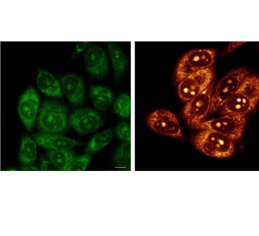
Application of RNA Imaging Probe 1c on live HeLa cells. Confocal fluorescence images of live Hela cells imaged with 20 μM methyl pyridinium indole (MPI) (green) ( lambda ex = 470 nm, lambda em = 525-555 nm) and 20 μM RNA Imaging Probe 1c (red) ( lambda ex = 550 nm, lambda em= 580-620 nm) for 30 min. Scale bar: 10 μm.
 View Larger
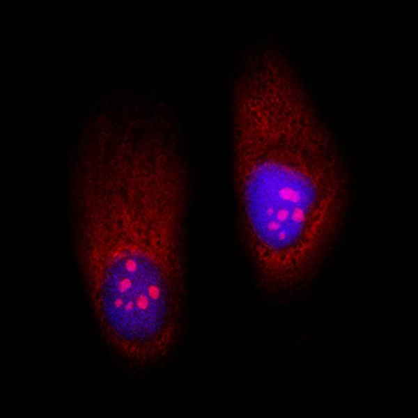
Application of RNA Imaging Probe 1c on PFA-fixed HeLa cells. Confocal fluorescence images of PFA-fixed HeLa cells incubated with 20 μM RNA Imaging Probe 1c (red) and 1 μg/mL Hoechst 33342 (blue) for 30 min.
 View Larger
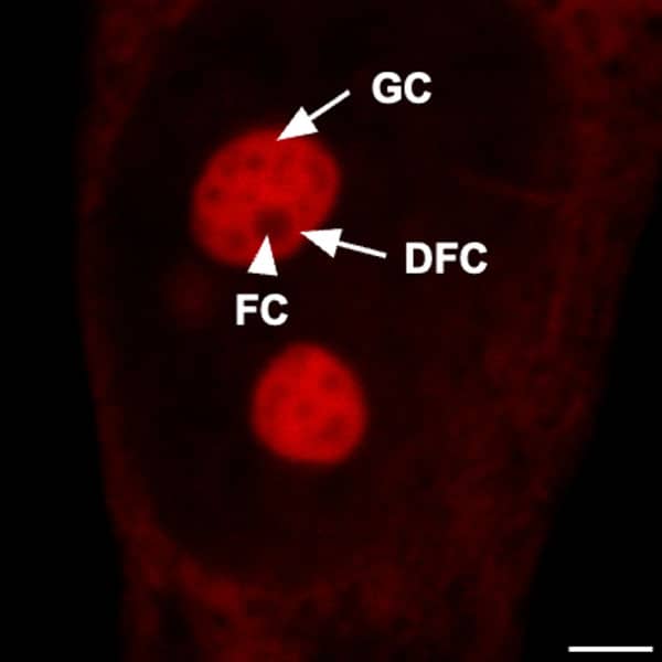
Application of RNA Imaging Probe 1c on HeLa cells. Zoomed-in confocal fluorescence image of HeLa cells stained with 20 μM RNA Imaging Probe 1c for 30 min. Image shows the sub-nucleolar components fibrillar center (FC) (indicated by the white arrowhead), dense fibrillar component (DFC), and outer granular component (GC) (indicated by the white arrows). Scale bar: 3 μm.
 View Larger
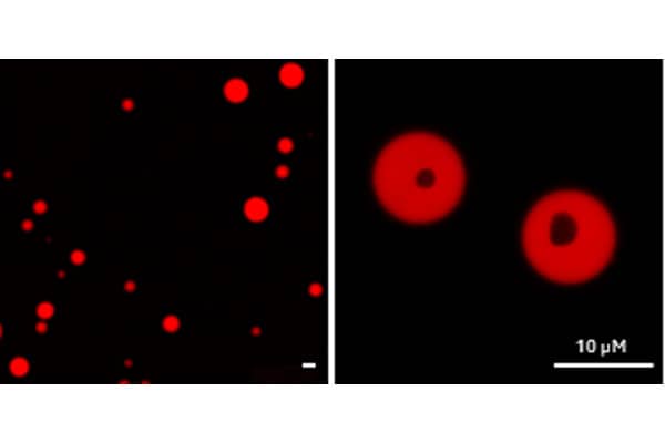
Yeast RNA and spermine labeled with RNA Imaging Probe 1c. Torula yeast RNA and spermine were incubated with RNA Imaging Probe 1c (10 μM). FLIM images show the formation of spherical coacervate droplets upon mixing the RNA and spermine. Image is enlarged by 1× (on the left) or by 8× (on the right). Scale bar: 10 μm.
Optical Data for RNA Imaging Probe 1c
| λabs | 556 nm |
|---|---|
| λem | 608 nm |
| Extinction Coefficient (ε) | 27500 M-1cm-1 |
| Quantum Yield (φ) | 0.49 |
| Cell Permeable | Yes |
Plan Your Experiments
Use our spectra viewer to interactively plan your experiments, assessing multiplexing options. View the excitation and emission spectra for our fluorescent dye range and other commonly used dyes.
Spectral ViewerTechnical Data
The technical data provided above is for guidance only.
For batch specific data refer to the Certificate of Analysis.
Tocris products are intended for laboratory research use only, unless stated otherwise.
Additional Information
Product Datasheets
FAQs
No product specific FAQs exist for this product, however you may
View all Small Molecule FAQsReviews for RNA Imaging Probe 1c
There are currently no reviews for this product. Be the first to review RNA Imaging Probe 1c and earn rewards!
Have you used RNA Imaging Probe 1c?
Submit a review and receive an Amazon gift card.
$25/€18/£15/$25CAN/¥75 Yuan/¥2500 Yen for a review with an image
$10/€7/£6/$10 CAD/¥70 Yuan/¥1110 Yen for a review without an image
