Mouse/Rat Cathepsin L Antibody Summary
Thr18-Asn334
Accession # P06797
Applications
Please Note: Optimal dilutions should be determined by each laboratory for each application. General Protocols are available in the Technical Information section on our website.
Scientific Data
 View Larger
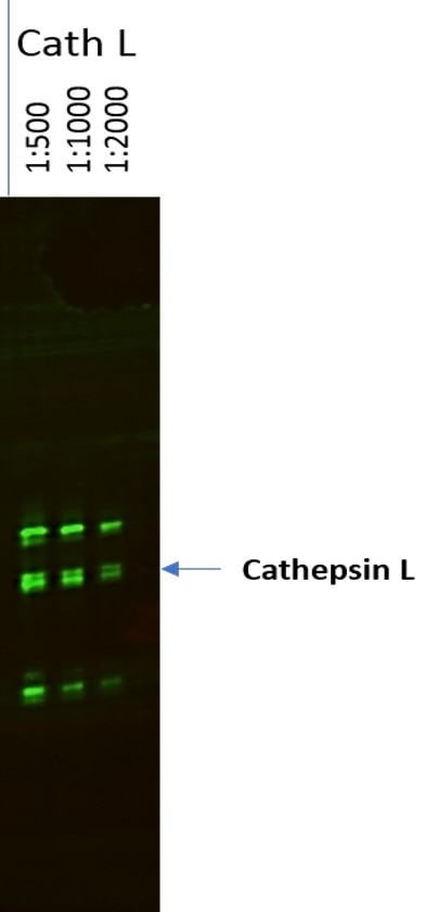
Detection of Mouse and Rat Cathepsin L by Western Blot. Western blot shows lysates of rat liver tissue, mouse liver tissue (wild type), and mouse liver tissue (knock out). PVDF membrane was probed with 1 µg/mL of Goat Anti-Mouse Cathepsin L Antigen Affinity-purified Polyclonal Antibody (Catalog # AF1515) followed by HRP-conjugated Anti-Goat IgG Secondary Antibody (HAF017). Specific bands were detected for Cathepsin L at approximately 22-38 kDa (as indicated). This experiment was conducted under reducing conditions and using Immunoblot Buffer Group 1.
 View Larger
Cathepsin L in Mouse Ovary. Cathepsin L was detected in perfusion fixed frozen sections of mouse ovary using 15 µg/mL Goat Anti-Mouse Cathepsin L Antigen Affinity-purified Polyclonal Antibody (Catalog # AF1515) overnight at 4 °C. Tissue was stained with the Anti-Goat HRP-DAB Cell & Tissue Staining Kit (brown; CTS008) and counterstained with hematoxylin (blue). View our protocol for Chromogenic IHC Staining of Frozen Tissue Sections.
 View Larger
Detection of Mouse Cathepsin L by Simple WesternTM. Simple Western lane view shows lysates of mouse liver tissue and HepG2 human hepatocellular carcinoma cell line, loaded at 0.2 mg/mL. Specific bands were detected for Cathepsin L at approximately 51 and 34 kDa (as indicated) using 10 µg/mL of Goat Anti-Mouse Cathepsin L Antigen Affinity-purified Polyclonal Antibody (Catalog # AF1515) followed by 1:50 dilution of HRP-conjugated Anti-Goat IgG Secondary Antibody (HAF109). This experiment was conducted under reducing conditions and using the 12-230 kDa separation system.
 View Larger
Cathepsin L in Mouse Thymus. Cathepsin L was detected in perfusion fixed frozen sections of mouse thymus using 15 µg/mL Goat Anti-Mouse Cathepsin L Antigen Affinity-purified Polyclonal Antibody (Catalog # AF1515) overnight at 4 °C. Tissue was stained with the Anti-Goat HRP-DAB Cell & Tissue Staining Kit (brown; CTS008) and counterstained with hematoxylin (blue). View our protocol for Chromogenic IHC Staining of Frozen Tissue Sections.
 View Larger
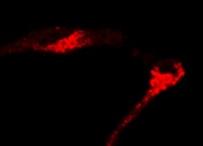
Detection of Mouse Cathepsin L by Immunocytochemistry/ Immunofluorescence Myc induces cathepsin L expression in beta-cells of pancreatic Islets.(A) Immunohistochemical analyses for CTS B, C, L or S expression (all in red) in combination with staining for the pan-leukocyte marker CD45 (green) in pancreatic islet tumors from the MycERTAM;Bcl-xL animals. Pancreata were harvested from the MycERTAM;Bcl-xL mice treated for 7 d with TAM (Myc-On, 7 days) or control vehicle in place of TAM (Myc-OFF). The islet area is indicated by dotted line. The asterisks indicate the area of tumor represented in the insets. The panels are representatives of at least three animals assayed at each data point, all immunohistochemical analyses were done in duplicate; eight randomized fields per analysis were examined. Scale bars, 100μm. (B) Immunohistochemical analysis for cathepsin L expression in beta-cells of pancreatic islets from MycERTAM;Bcl-xL animals identified by insulin expression. Pancreata were collected from the animals described above. Scale bars represent 25μm. The panels are representatives of three animals assayed at each data point, all immunohistochemical analyses were done in duplicate; ten randomized fields per analysis were examined. Image collected and cropped by CiteAb from the following open publication (https://dx.plos.org/10.1371/journal.pone.0120348), licensed under a CC-BY license. Not internally tested by R&D Systems.
Preparation and Storage
- 12 months from date of receipt, -20 to -70 °C as supplied.
- 1 month, 2 to 8 °C under sterile conditions after reconstitution.
- 6 months, -20 to -70 °C under sterile conditions after reconstitution.
Background: Cathepsin L
Cathepsin L is a lysosomal cysteine protease expressed in most eukaryotic cells. Cathepsin L is known to hydrolyze a number of proteins, including the proform of urokinase-type plasminogen activator, which is activated by Cathepsin L cleavage (1). Cathepsin L has also been shown to proteolytically inactivate alpha 1-antitrypsin and secretory leucoprotease inhibitor, two major protease inhibitors of the respiratory tract (2). These observations, combined with the demonstration of increased Cathepsin L activity in the epithelial lining fluid of the lungs of emphysema patients, have led to the suggestion that the enzyme may be involved in the progression of this disease. Cathepsin L has also been identified as a major excreted protein of transformed fibroblasts, indicating the enzyme could be involved in malignant tumor growth (3). In Cathepsin L-deficient mice, it appears to play a critical role in cardiac morphology and function, epidermal homeostasis, regulation of the hair cycle, and MHC class II-mediated antigen presentation in cortical epithelial cells of the thymus (4, 5). Mouse Cathepsin L is synthesized as a 334 amino acid precursor with a signal peptide (residues 1-17), a pro region (residues 18-113), and a mature chain (residues 114-334).
- Goretzki, L. et al. (1992) FEBS Lett. 297:112.
- Taggart, C.C. et al. (2001) J. Biol. Chem. 276:33345.
- Gottesman, M.M. and F. Cabral (1981) Biochemistry 20:1659.
- Stypmann, J. et al. (2002) Proc. Natl. Acad. Sci. USA 99: 6234.
- Reinheckel, T. et al. (2001) Biol. Chem. 382:735.
Product Datasheets
Citations for Mouse/Rat Cathepsin L Antibody
R&D Systems personnel manually curate a database that contains references using R&D Systems products. The data collected includes not only links to publications in PubMed, but also provides information about sample types, species, and experimental conditions.
47
Citations: Showing 1 - 10
Filter your results:
Filter by:
-
Out-of-frame start codons prevent translation of truncated nucleo-cytosolic cathepsin L in vivo
Authors: Martina Tholen, Larissa E. Hillebrand, Stefan Tholen, Oliver Sedelmeier, Sebastian J. Arnold, Thomas Reinheckel
Nature Communications
-
(-)-Oleocanthal and (-)-oleocanthal-rich olive oils induce lysosomal membrane permeabilization in cancer cells
Authors: Limor Goren, George Zhang, Susmita Kaushik, Paul A. S. Breslin, Yi-Chieh Nancy Du, David A. Foster
PLOS ONE
-
Loss of TMEM106B and PGRN leads to severe lysosomal abnormalities and neurodegeneration in mice
Authors: Feng T, Mai S, Roscoe JM et al.
EMBO Rep.
-
Identification of murine gammaherpesvirus 68 miRNA-mRNA hybrids reveals miRNA target conservation among gammaherpesviruses including host translation and protein modification machinery
Authors: Bullard WL, Kara M, Gay LA et al.
PLoS Pathog.
-
Loss of TMEM106B Ameliorates Lysosomal and Frontotemporal Dementia-Related Phenotypes in Progranulin-Deficient Mice
Authors: Zoe A. Klein, Hideyuki Takahashi, Mengxiao Ma, Massimiliano Stagi, Melissa Zhou, TuKiet T. Lam et al.
Neuron
-
cPLA2 activation contributes to lysosomal defects leading to impairment of autophagy after spinal cord injury
Authors: Y Li, JW Jones, H M C Choi, C Sarkar, MA Kane, EY Koh, MM Lipinski, J Wu
Cell Death Dis, 2019-07-11;10(7):531.
-
A multifaceted role of progranulin in regulating amyloid-beta dynamics and responses
Authors: Du H, Wong MY, Zhang T et al.
Life science alliance
-
Development of Activity-Based Probes for Cathepsin X
Authors: Margot G. Paulick, Matthew Bogyo
ACS Chemical Biology
-
Elevated mRNA expression and defective processing of cathepsin D in HeLa cells lacking the mannose 6-phosphate pathway
Authors: L Liu, B Doray
FEBS Open Bio, 2021-05-05;0(0):.
-
Cathepsin L Regulates Metabolic Networks Controlling Rapid Cell Growth and Proliferation
Authors: Weiss Sadan, T;Itzhak, G;Kaschani, F;Yu, Z;Mahameed, M;Anaki, A;Ben-Nun, Y;Merquiol, E;Tirosh, B;Kessler, B;Kaiser, M;Blum, G;
Mol. Cell Proteomics
-
Investigations on Primary Cilia of Nthy-ori 3-1 Cells upon Cysteine Cathepsin Inhibition or Thyrotropin Stimulation
Authors: Do?ru, AG;Rehders, M;Brix, K;
International journal of molecular sciences
Species: Human
Sample Types: Cell Lysates
Applications: Western Blot -
Elamipretide alleviates pyroptosis in traumatically injured spinal cord by inhibiting cPLA2-induced lysosomal membrane permeabilization
Authors: H Zhang, Y Chen, F Li, C Wu, W Cai, H Ye, H Su, M He, L Yang, X Wang, K Zhou, W Ni
Journal of Neuroinflammation, 2023-01-07;20(1):6.
Species: Mouse
Sample Types: Tissue Homogenates, Whole Tissue
Applications: IHC, Western Blot -
Spatiotemporal organisation of protein processing in the kidney
Authors: M Polesel, M Kaminska, D Haenni, M Bugarski, C Schuh, N Jankovic, A Kaech, JM Mateos, M Berquez, AM Hall
Nature Communications, 2022-09-29;13(1):5732.
Species: Mouse
Sample Types: Whole Tissue
Applications: IHC -
Direct control of lysosomal catabolic activity by mTORC1 through regulation of V-ATPase assembly
Authors: E Ratto, SR Chowdhury, NS Siefert, M Schneider, M Wittmann, D Helm, W Palm
Nature Communications, 2022-08-17;13(1):4848.
Species: Mouse
Sample Types: Cell Lysates
Applications: Western Blot -
Cas13d knockdown of lung protease Ctsl prevents and treats SARS-CoV-2 infection
Authors: Z Cui, C Zeng, F Huang, F Yuan, J Yan, Y Zhao, Y Zhou, W Hankey, VX Jin, J Huang, HF Staats, JI Everitt, GD Sempowski, H Wang, Y Dong, SL Liu, Q Wang
Oncogene, 2022-07-25;0(0):.
Species: Mouse
Sample Types: Tissue Homogenates, Whole Tissue
Applications: IHC, Western Blot -
TMEM106B deficiency impairs cerebellar myelination and synaptic integrity with Purkinje cell loss
Authors: T Feng, L Luan, II Katz, M Ullah, VM Van Deerli, JQ Trojanowsk, EB Lee, F Hu
Acta neuropathologica communications, 2022-03-14;10(1):33.
Species: Mouse
Sample Types: Tissue Homogenates
Applications: Western Blot -
Translation Inhibitors Activate Autophagy Master Regulators TFEB and TFE3
Authors: TT Dang, SH Back
International Journal of Molecular Sciences, 2021-11-08;22(21):.
Species: Mouse
Sample Types: Cell Lysates
Applications: Western Blots -
Chitinase 3-like-1 is a Therapeutic Target That Mediates the Effects of Aging in COVID-19
Authors: S Kamle, B Ma, CH He, B Akosman, Y Zhou, CM Lee, WS El-Deiry, K Huntington, O Liang, J Machan, MJ Kang, HJ Shin, E Mizoguchi, CG Lee, JA Elias
bioRxiv : the preprint server for biology, 2021-02-16;0(0):.
Species: Mouse
Sample Types: Tissue Homogenates, Whole Tissue
Applications: IHC, Western Blot -
Cathepsin L regulates pathogenicCD4 T cells in experimental autoimmune encephalomyelitis
Authors: M Shibamura-, K Yuki, L Hou
International immunopharmacology, 2021-02-01;93(0):107425.
Species: Mouse
Sample Types: Cell Lysates
Applications: Western Blot -
Imbalanced cellular metabolism compromises cartilage homeostasis and joint function in a mouse model of mucolipidosis type III gamma
Authors: LM Westermann, L Fleischhau, J Vogel, Z Jenei-Lanz, N Floriano L, L Schau, F Morellini, A Baranowsky, TA Yorgan, G Di Lorenzo, M Schweizer, B de Souza P, NR Guarany, F Sperb-Ludw, F Visioli, T Oliveira S, J Soul, G Hendrickx, JS Wiegert, IVD Schwartz, H Clausen-Sc, F Zaucke, T Schinke, S Pohl, T Danyukova
Dis Model Mech, 2020-11-18;0(0):.
Species: Mouse
Sample Types: Cell Lysates
Applications: Western Blot -
Cathepsin D deficiency in mammary epithelium transiently stalls breast cancer by interference with mTORC1 signaling
Authors: S Ketterer, J Mitschke, A Ketscher, M Schlimpert, W Reichardt, N Baeuerle, ME Hess, P Metzger, M Boerries, C Peters, B Kammerer, T Brummer, F Steinberg, T Reinheckel
Nat Commun, 2020-10-12;11(1):5133.
Species: Mouse
Sample Types: Cell Lysates
Applications: Western Blot -
Conditional Gene Targeting Reveals Cell Type-Specific Roles of the Lysosomal Protease Cathepsin L in Mammary Tumor Progression
Authors: MA Parigiani, A Ketscher, S Timme, P Bronsert, M Schlimpert, B Kammerer, A Jacquel, P Chaintreui, T Reinheckel
Cancers (Basel), 2020-07-22;12(8):.
Species: Mouse
Sample Types: Cell Culture Lysates
Applications: Western Blot -
PLA2G4A/cPLA2-mediated lysosomal membrane damage leads to inhibition of autophagy and neurodegeneration after brain trauma
Authors: C Sarkar, JW Jones, N Hegdekar, JA Thayer, A Kumar, AI Faden, MA Kane, MM Lipinski
Autophagy, 2019-06-25;0(0):1-20.
Species: Mouse
Sample Types: Whole Cells, Whole Tissue
Applications: ICC, IHC -
Sequential, but not Concurrent, Incubation of Cathepsin K and L with Type I Collagen Results in Extended Proteolysis
Authors: AN Parks, J Nahata, NE Edouard, JS Temenoff, MO Platt
Sci Rep, 2019-04-01;9(1):5399.
Species: Human
Sample Types: Recombinant Protein
Applications: Western Blot -
Early lysosomal maturation deficits in microglia triggers enhanced lysosomal activity in other brain cells of progranulin knockout mice
Authors: JK Götzl, AV Colombo, K Fellerer, A Reifschnei, G Werner, S Tahirovic, C Haass, A Capell
Mol Neurodegener, 2018-09-04;13(1):48.
Species: Mouse
Sample Types: Cell Lysates
Applications: Western Blot -
Stat3 mediated alterations in lysosomal membrane protein composition
Authors: B Lloyd-Lewi, CC Krueger, TJ Sargeant, ME D'Angelo, MJ Deery, R Feret, JA Howard, KS Lilley, CJ Watson
J. Biol. Chem., 2018-01-17;0(0):.
Species: Mouse
Sample Types: Cell Culture Supernates
Applications: Western Blot -
Investigating the Life Expectancy and Proteolytic Degradation of Engineered Skeletal Muscle Biological Machines
Authors: C Cvetkovic, MC Ferrall-Fa, E Ko, L Grant, H Kong, MO Platt, R Bashir
Sci Rep, 2017-06-19;7(1):3775.
Species: Mouse
Sample Types: Tissue Homogenates
Applications: Western Blot -
The Role of Heparanase in the Pathogenesis of Acute Pancreatitis: A Potential Therapeutic Target
Authors: I Khamaysi, P Singh, S Nasser, H Awad, Y Chowers, E Sabo, E Hammond, I Gralnek, I Minkov, A Noseda, N Ilan, I Vlodavsky, Z Abassi
Sci Rep, 2017-04-06;7(1):715.
Species: Mouse
Sample Types: Whole Tissue
Applications: IHC -
Direct Observation of Enhanced Nitric Oxide in a Murine Model of Diabetic Nephropathy
Authors: MG Boels, EE van Faasse, MC Avramut, J van der Vl, BM van den Be, TJ Rabelink
PLoS ONE, 2017-01-19;12(1):e0170065.
Species: Mouse
Sample Types: Whole Tissue
Applications: IHC-P -
Deficiency for the cysteine protease cathepsin L impairs Myc-induced tumorigenesis in a mouse model of pancreatic neuroendocrine cancer.
Authors: Brindle N, Joyce J, Rostker F, Lawlor E, Swigart-Brown L, Evan G, Hanahan D, Shchors K
PLoS ONE, 2015-04-30;10(4):e0120348.
Species: Mouse
Sample Types: Whole Tissue
Applications: IHC -
Vacuolar ATPase in phagosome-lysosome fusion.
Authors: Kissing S, Hermsen C, Repnik U, Nesset C, von Bargen K, Griffiths G, Ichihara A, Lee B, Schwake M, De Brabander J, Haas A, Saftig P
J Biol Chem, 2015-04-22;290(22):14166-80.
Species: Mouse
Sample Types: Cell Lysates
Applications: Western Blot -
Lysosomal protein turnover contributes to the acquisition of TGFbeta-1 induced invasive properties of mammary cancer cells.
Authors: Kern U, Wischnewski V, Biniossek M, Schilling O, Reinheckel T
Mol Cancer, 2015-02-15;14(0):39.
Species: Mouse
Sample Types: Cell Lysates
Applications: Western Blot -
The PI3K regulatory subunits p55alpha and p50alpha regulate cell death in vivo.
Authors: Pensa S, Neoh K, Resemann H, Kreuzaler P, Abell K, Clarke N, Reinheckel T, Kahn C, Watson C
Cell Death Differ, 2014-06-06;21(9):1442-50.
Species: Mouse
Sample Types: Cell Lysates
Applications: Western Blot -
Biogenesis and proteolytic processing of lysosomal DNase II.
Authors: Ohkouchi, Susumu, Shibata, Masahiro, Sasaki, Mitsuho, Koike, Masato, Safig, Paul, Peters, Christop, Nagata, Shigekaz, Uchiyama, Yasuo
PLoS ONE, 2013-03-13;8(3):e59148.
Species: Mouse
Sample Types: Whole Tissue
Applications: IHC -
Macrophages and cathepsin proteases blunt chemotherapeutic response in breast cancer.
Authors: Shree T, Olson OC, Elie BT
Genes Dev., 2011-12-01;25(23):2465-79.
Species: Mouse
Sample Types: Tissue Homogenates
Applications: Western Blot -
Major Role of Cathepsin L for Producing the Peptide Hormones ACTH, beta-Endorphin, and alpha-MSH, Illustrated by Protease Gene Knockout and Expression.
Authors: Funkelstein L, Toneff T, Mosier C, Hwang SR, Beuschlein F, Lichtenauer UD, Reinheckel T, Peters C, Hook V
J. Biol. Chem., 2008-10-10;283(51):35652-9.
Species: Mouse
Sample Types: Whole Tissue
Applications: IHC-Fr -
The impact of microRNAs on protein output.
Authors: Baek D, Villen J, Shin C, Camargo FD, Gygi SP, Bartel DP
Nature, 2008-07-30;455(7209):64-71.
Species: Mouse
Sample Types: Cell Lysates
Applications: Western Blot -
Maternal Transmission of a Humanised Igf2r Allele Results in an Igf2 Dependent Hypomorphic and Non-Viable Growth Phenotype
Authors: Jennifer Hughes, Susana Frago, Claudia Bühnemann, Emma J. Carter, A. Bassim Hassan
PLoS ONE
-
GCN2 adapts protein synthesis to scavenging-dependent growth
Authors: Michel Nofal, Tim Wang, Lifeng Yang, Connor S.R. Jankowski, Sophia Hsin-Jung Hsin-Jung Li, Seunghun Han et al.
Cell Systems
-
CCT complex restricts neuropathogenic protein aggregation via autophagy
Nat Commun, 2016-12-08;7(0):13821.
-
Critical Role of Cathepsin L/V in Regulating Endothelial Cell Senescence
Authors: Chan Li, Zhaoya Liu, Mengshi Chen, Liyang Zhang, Ruizheng Shi, Hua Zhong
Biology (Basel)
-
The protease cathepsin L regulates Th17 cell differentiation
Authors: Lifei Hou, Jessica Cooley, Richard Swanson, Poh Chee Ong, Robert N. Pike, Matthew Bogyo et al.
Journal of Autoimmunity
-
Chitinase 3-like-1 is a therapeutic target that mediates the effects of aging in COVID-19
Authors: Suchitra Kamle, Bing Ma, Chuan Hua He, Bedia Akosman, Yang Zhou, Chang-Min Lee et al.
JCI Insight
-
Stat3 controls cell death during mammary gland involution by regulating uptake of milk fat globules and lysosomal membrane permeabilization
Authors: Timothy J. Sargeant, Bethan Lloyd-Lewis, Henrike K. Resemann, Antonio Ramos-Montoya, Jeremy Skepper, Christine J. Watson
Nature Cell Biology
-
Loss of TMEM 106B potentiates lysosomal and FTLD ‐like pathology in progranulin‐deficient mice
Authors: Georg Werner, Markus Damme, Martin Schludi, Johannes Gnörich, Karin Wind, Katrin Fellerer et al.
EMBO reports
-
Acute, Delayed and Chronic Remote Ischemic Conditioning Is Associated with Downregulation of mTOR and Enhanced Autophagy Signaling.
Authors: Rohailla S, Clarizia N, Sourour M et al.
PLoS OnE.
-
Mice Hypomorphic for Keap1, a Negative Regulator of the Nrf2 Antioxidant Response, Show Age-Dependent Diffuse Goiter with Elevated Thyrotropin Levels
Authors: Panos G. Ziros, Cédric O. Renaud, Dionysios V. Chartoumpekis, Massimo Bongiovanni, Ioannis G. Habeos, Xiao-Hui Liao et al.
Thyroid
FAQs
No product specific FAQs exist for this product, however you may
View all Antibody FAQsReviews for Mouse/Rat Cathepsin L Antibody
Average Rating: 5 (Based on 3 Reviews)
Have you used Mouse/Rat Cathepsin L Antibody?
Submit a review and receive an Amazon gift card.
$25/€18/£15/$25CAN/¥75 Yuan/¥2500 Yen for a review with an image
$10/€7/£6/$10 CAD/¥70 Yuan/¥1110 Yen for a review without an image
Filter by:


