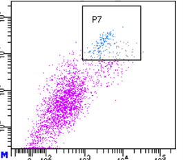
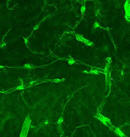
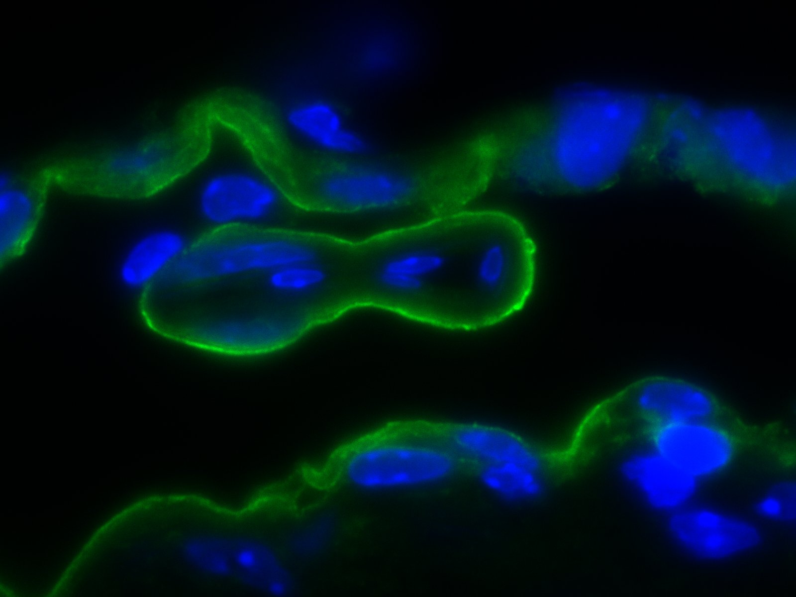
Mouse PDGF R beta Antibody Summary
Leu32-Lys530
Accession # P05622
*Small pack size (-SP) is supplied either lyophilized or as a 0.2 µm filtered solution in PBS.
Applications
Please Note: Optimal dilutions should be determined by each laboratory for each application. General Protocols are available in the Technical Information section on our website.
Scientific Data
 View Larger
Detection of Rat and Mouse PDGF R beta by Western Blot. Western blot shows lysates of rat spinal cord tissue, rat brain tissue, and mouse brain tissue. PVDF membrane was probed with 0.25 µg/mL of Goat Anti-Mouse PDGF R beta Antigen Affinity-purified Polyclonal Antibody (Catalog # AF1042) followed by HRP-conjugated Anti-Goat IgG Secondary Antibody (Catalog # HAF019). A specific band was detected for PDGF R beta at approximately 190 kDa (as indicated). This experiment was conducted under reducing conditions and using Immunoblot Buffer Group 1.
 View Larger
PDGF R beta in Mouse Embryonic Spinal Cord. PDGF R beta was detected in immersion fixed frozen sections of mouse embryo spinal cord using 5 µg/mL Goat Anti-Mouse PDGF R beta Antigen Affinity-purified Polyclonal Antibody (Catalog # AF1042) overnight at 4 °C. Tissue was stained with the Anti-Goat HRP-DAB Cell & Tissue Staining Kit (brown; Catalog # CTS008) and counterstained with hematoxylin (blue). View our protocol for Chromogenic IHC Staining of Frozen Tissue Sections.
 View Larger
Detection of Mouse PDGF R beta by Immunohistochemistry Expression of sFlt1 in pericytes at the angiogenic front. a Maximum intensity projections of confocal images from P6 retinas of the Hey1-GFP transgenic reporter mouse model stained for GFP (green), PDGFR beta + (white) and IB4 (red). Images of the first row show enrichment of Hey1-GFP+, PDGFR beta + perivascular cells in the angiogenic front in comparison to mural cells covering the remodeling central plexus around veins (middle row) and arteries (bottom row). Note strong expression of Hey1-GFP reporter in arterial ECs (bottom row). Scale bar, 50 µm. b, Quantitation of Pdgfrb expression by qPCR in P6 PDGFR beta + retinal pericytes sorted based on GFP expression in comparison to whole-retina single-cell suspension (input). Note significant enrichment of Pdgfrb in both (GFP+ and GFP−) pericyte fractions and higher expression in the Hey1-GFP+ subset. Error bars, s.e.m. p-values, Kruskal–Wallis and Dunn’s multiple comparison test. NS, not statistically significant. c Quantitation of sFlt1 expression by qPCR in sorted P6 retinal pericytes in comparison to whole-retina single-cell suspension (input). Note significant enrichment of sFlt1 expression in Hey1-GFP+ pericytes in comparison to input and GFP- pericytes. Error bars, s.e.m. p-values, one-way ANOVA and Tukey’s multiple comparison test. NS, not statistically significant Image collected and cropped by CiteAb from the following publication (https://pubmed.ncbi.nlm.nih.gov/29146905), licensed under a CC-BY license. Not internally tested by R&D Systems.
 View Larger
Detection of Mouse PDGF R beta by Immunocytochemistry/Immunofluorescence VCAM1 is involved in the regulation of proliferation and morphological changes in oligodendrocytes.(a) Tissue lysates from 7-day-old NG2-Cre-driven VCAM1 conditional knockout (VCAM1fl/fl; Ng2-Cre) or control (Ctrl) mouse spinal cords or 11-day-old whole brains were immunoblotted with an antibody against MBP, CNPase, VCAM1 or actin. Data are representative of three experiments. (b–d) Antibodies against Ki67 (green) and Olig2 (red) were used for co-staining in 2-day-old spinal cord cross sections. Data are representative. The scale bars indicate 200 μm. The number of Olig2+ cells per one square millimetre was counted. Data were evaluated using Student’s t-test (**P=0.000528; n=19 slices of two independent experiments). The percentage of Ki67+ cells among the Olig2+ cells is shown. Data were evaluated using Student’s t-test (**P=0.00807; n=6 slices of two independent experiments). (e) Antibodies against Ki67 and NG2 were used for co-staining in 2-day-old spinal cord. The percentage of Ki67+ cells among the NG2+ cells per one square millimetre is shown. Data were evaluated using Student’s t-test (*P=0.0275; n=6 slices of two independent experiments). (f–h) Knock-down efficiencies of shRNAs were confirmed by immunoblotting. Data are representative of three experiments. Oligodendrocytes were transfected with VCAM1 shRNA or control, co-cultured with neurons for 2 days, and co-stained with antibodies against NF (green) and NG2 (red). Magnified images of the dotted boxes (i, attached; ii, aligned) are shown below. Data are representative. Scale bar, 100 μm. The percentage of NG2+ cells whose process tips or cell bodies were attached to axons or aligned along axons is shown. Data were evaluated using Student’s t-test (**P=0.00109 (attached cells) or P=0.00982 (aligned cells); n=6 areas of three experiments). (i,k) Oligodendrocytes were cultured on dishes coated with or without recombinant alpha 4 beta 1 integrin in a growth medium containing PDGF and bFGF. Cells were co-stained with an anti-MBP antibody (red) and DAPI (blue) (a) or an anti-PDGFR alpha antibody (green) (b). Data are representative. Scale bar, 100 μm. The percentage of MBP+ cells is shown. Data were evaluated using Student’s t-test (**P=1.14E−10; n=6 areas of two independent experiments). (j,l) Oligodendrocytes transfected with VCAM1#1 or control shRNA were cultured on recombinant alpha 4 beta 1 integrin-coated dishes in the differentiation medium and co-stained with an anti-MBP antibody (red) and DAPI (blue). Data are representative. Scale bar, 100 μm. The percentage of MBP+ cells is shown. Data were evaluated using Student’s t-test (**P=1.82E−21; n=6 areas of two independent experiments). (m–o) Antibodies against Ki67 (green) and O4 (red) were used for co-staining in 7-day-old spinal cord. DAPI staining is also shown. Data are representative. Scale bar, 100 μm. The number of O4+ cells was counted. Data were evaluated using Student’s t-test (**P=5.87E−05 (P7) or P=5.87E−06 (P14); n=12 slices of two independent experiments). The percentage of Ki67− or Ki67+ cells among the O4+ cells is shown. Data were evaluated using Student’s t-test (**P=0.00344 (P7, Ki67−), 0.00344 (P7, Ki67+) or 0.000580 (P14, Ki67+) *P=0.0494 (P14, Ki67−); n=12 slices of two independent experiments). Image collected and cropped by CiteAb from the following publication (https://pubmed.ncbi.nlm.nih.gov/27876794), licensed under a CC-BY license. Not internally tested by R&D Systems.
 View Larger
Detection of Mouse PDGF R beta by Immunohistochemistry Vascular alterations after intraocular VEGF-A injection. a Morphology of IB4-stained P6 wild-type retinal vessels at 4 h after administration of human VEGF-A165 (0.5 µl at a concentration of 5 μg μl−1). Note blunt appearance of the vessel front after VEGF-A injection but not for vehicle (PBS) control. Scale bar, 200 µm. b Quantitation of sprouts and filopodia at the front of the P6 vessel plexus after injection of VEGF-A165 or vehicle control. Error bars, s.e.m. p-values, Student’s t-test. c PDGFR beta + (green) pericytes are unaffected by short-term VEGF-A administration, whereas VEGFR2 immunosignals (white) are increased in IB4+ (red) ECs (arrowheads). Images shown correspond to insets in a. Scale bar, 100 µm. d Quantitation of VEGFR2 immunosignals intensity in the peripheral plexus of P6 retinas after injection of VEGF-A165 or vehicle control. Error bars, s.e.m. p-values, Student’s t-test. e Confocal images showing increased Esm1 immunostaining (white) in IB4+ (red) ECs in the peripheral plexus (arrowheads) after VEGF-A injection in P6 pups. Scale bar, 200 µm. f VEGF-A165 injection-mediated increase of Esm1 immunosignals (normalized to IB4+ EC area) in the peripheral capillary plexus but not at the edge of the angiogenic front in comparison to PBS-injected controls at P6. Error bars, s.e.m. p-values, Student’s t-test. NS, not statistically significant. g Short-term VEGF-A165 administration leads to clustering of Erg1+ (green) and IB4+ (red) ECs, as indicated, in thick sprout-like structures of P6 retinas. Panels in the center and on the right (scale bar, 20 µm) show higher magnification of the insets on the left (scale bar, 100 µm). Dashed lines in panels on the right outline IB4+ vessels. h Quantitation of EC density in the leading front vessel and emerging sprouts of the P6 angiogenic front after injection of VEGF-A165 or vehicle control. Error bars, s.e.m. p-values, Student’s t-test Image collected and cropped by CiteAb from the following publication (https://pubmed.ncbi.nlm.nih.gov/29146905), licensed under a CC-BY license. Not internally tested by R&D Systems.
 View Larger
Detection of PDGF R beta in Mouse Embryo Brain. Formalin-fixed paraffin-embedded tissue sections of mouse embryo were probed for PDGFRb mRNA (ACD RNAScope Probe, catalog #411388; Fast Red chromogen, ACD catalog # 322750). Adjacent tissue section was processed for immunohistochemistry using goat anti-mouse PDGFRb polyclonal antibody (R&D Systems catalog # AF1042) at 1.7ug/mL with overnight incubation at 4 degrees Celsius followed by incubation with anti-goat IgG VisUCyte HRP Polymer Antibody (Catalog # VC004) and DAB chromogen (yellow-brown). Tissue was counterstained with hematoxylin (blue). Specific staining was localized to developing vasculature.
 View Larger
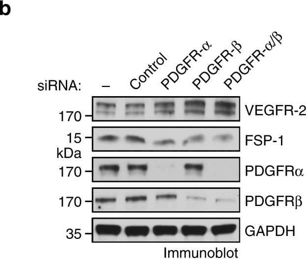
Detection of Mouse PDGF R beta by Western Blot PDGF autocrine loop is critical for VEGFR-2 down-expression and anti-VEGF resistance in GBM-associated ECs. a GBM tumor-derived ECs were treated with control IgG or antibody against PDGF-AA or PDGF-BB. Cell lysates were immunoblotted. b, c GBM tumor-derived ECs were transfected with control scrambled siRNA or siRNA targeting PDGFR-alpha and PDGFR-beta. b Cell lysates were immunoblotted. c Cell proliferation was determined (n = 3, mean ± SEM). d GBM tumor-derived ECs were treated with Ki8751 and crenolanib at different doses. Cell proliferation was determined 4 days after treatment. Inhibition rates were calculated and expressed as % of control cells Image collected and cropped by CiteAb from the following open publication (https://pubmed.ncbi.nlm.nih.gov/30150753), licensed under a CC-BY license. Not internally tested by R&D Systems.
 View Larger
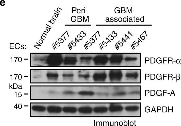
Detection of Human PDGF R beta by Western Blot PDGF induces downregulation of VEGFR-2 expression in ECs. a, b Normal human brain microvascular ECs (#1 and #2 from adult brain and #3 from fetal brain) were treated with glioma-conditioned medium (glioma-CM). RNA was isolated and subjected to transcriptome analysis by RNA deep sequencing (RNA-seq). Left, heat map for expression of VEGF receptors. Right, fold changes of VEGFR-1, VEGFR-2, and VEGFR-3 (n = 3, mean ± SEM). b Shown are FPKM values of FSP-1 (n = 3). c Normal brain ECs were treated with glioma-CM or control normal medium. Cell lysates were immunoblotted. d Gene set analysis of upregulated pathways/genes identified by RNA-seq in glioma-CM-treated ECs. e ECs were isolated from GBM tumors or peri-tumor tissues of human patients or normal brains. Cell lysates were immunoblotted. Note: the lyates were also immunoblotted in Fig. 1c, and the same blot for GAPDH was shown. f Normal brain ECs were treated with 100 ng/ml PDGF-AA, PDGF-AB, or PDGF-BB. Cell lysates were immunoblotted Image collected and cropped by CiteAb from the following open publication (https://pubmed.ncbi.nlm.nih.gov/30150753), licensed under a CC-BY license. Not internally tested by R&D Systems.
Preparation and Storage
- 12 months from date of receipt, -20 to -70 °C as supplied.
- 1 month, 2 to 8 °C under sterile conditions after reconstitution.
- 6 months, -20 to -70 °C under sterile conditions after reconstitution.
Background: PDGF R beta
The platelet-derived growth factor (PDGF) family consists of proteins derived from four genes (PDGF-A, -B, -C, and -D) that form disulfide-linked homodimers (PDGF-AA, -BB, -CC, and -DD) and a heterodimer (PDGF-AB) (1, 2). These proteins regulate diverse cellular functions by binding to and inducing the homo- or hetero-dimerization of two receptors (PDGF R alpha and R beta ). Whereas alpha / alpha homo-dimerization is induced by PDGF-AA, -BB, -CC, and -AB, alpha / beta hetero-dimerization is induced by PDGF-AB, -BB, -CC, and -DD, and beta / beta homo-dimerization is induced only by PDGF-BB, and -DD (1 - 4). Both PDGF R alpha and R beta are members of the class III subfamily of receptor tyrosine kinases (RTK) that also includes the receptors for M-CSF, SCF and Flt3-ligand. All class III RTKs are characterized by the presence of five immunoglobulin-like domains in their extracellular region and a split kinase domain in their intracellular region. Ligand-induced receptor dimerization results in autophosphorylation in trans resulting in the activation of several intracellular signaling pathways that can lead to cell proliferation, cell survival, cytoskeletal rearrangement, and cell migration. Many cell types, including fibroblasts and smooth muscle cells, express both the alpha and beta receptors. Others have only the alpha receptors (oligodendrocyte progenitor cells, mesothelial cells, liver sinusoidal endothelial cells, astrocytes, platelets and megakaryocytes) or only the beta receptors (myoblasts, capillary endothelial cells, pericytes, T cells, myeloid hematopoietic cells and macrophages). A soluble PDGF R alpha has been detected in normal human plasma and serum as well as in the conditioned medium of the human osteosarcoma cell line MG-63 (5). Both the recombinant mouse and human soluble PDGF R alpha bind PDGF with high affinity and are potent PDGF antagonists.
- Betshotz, C. et al. (2001) BioEssays 23:494.
- Ostman, A. and A.H. Heldin (2001) Advances in Cancer Research 80:1.
- Gilbertson, D. et al. (2001) J. Biol. Chem. 276:27406.
- LaRochells, W.J. et al. (2001) Nature Cell Biol. 3:517.
- Tiesman, J. and C.E. Hart (1993) J. Biol. Chem. 5:9621.
Product Datasheets
Citations for Mouse PDGF R beta Antibody
R&D Systems personnel manually curate a database that contains references using R&D Systems products. The data collected includes not only links to publications in PubMed, but also provides information about sample types, species, and experimental conditions.
110
Citations: Showing 1 - 10
Filter your results:
Filter by:
-
Imatinib inhibits pericyte-fibroblast transition and inflammation and promotes axon regeneration by blocking the PDGF-BB/PDGFR? pathway in spinal cord injury
Authors: Yao F, Luo Y, Liu YC et al.
Inflammation and Regeneration
-
Weibel-Palade Bodies Orchestrate Pericytes During Angiogenesis
Authors: Mélissande Cossutta, Marie Darche, Gilles Carpentier, Claire Houppe, Matteo Ponzo, Fabio Raineri et al.
Arteriosclerosis, Thrombosis, and Vascular Biology
-
Dysfunction of Mouse Cerebral Arteries during Early Aging
Authors: Matilde Balbi, Mitrajit Ghosh, Thomas A Longden, Max Jativa Jativa Vega, Benno Gesierich, Farida Hellal et al.
Journal of Cerebral Blood Flow & Metabolism
-
A distant, cis-acting enhancer drives induction of Arf by Tgf beta in the developing eye
Authors: Yanbin Zheng, Caitlin Devitt, Jing Liu, Jie Mei, Stephen X. Skapek
Developmental Biology
-
Delayed Effects of Acute Reperfusion on Vascular Remodeling and Late-Phase Functional Recovery After Stroke
Authors: Violeta Durán-Laforet, David Fernández-López, Alicia García-Culebras, Juan González-Hijón, Ana Moraga, Sara Palma-Tortosa et al.
Frontiers in Neuroscience
-
Low-dose sodium-glucose cotransporter 2 inhibitor ameliorates ischemic brain injury in mice through pericyte protection without glucose-lowering effects
Authors: Masamitsu Takashima, Kuniyuki Nakamura, Takuya Kiyohara, Yoshinobu Wakisaka, Masaoki Hidaka, Hayato Takaki et al.
Communications Biology
-
A functional circuit formed by the autonomic nerves and myofibroblasts controls mammalian alveolar formation for gas exchange
Authors: Kuan Zhang, Erica Yao, Shao-An Wang, Ethan Chuang, Julia Wong, Liliana Minichiello et al.
Developmental Cell
-
Effects of Angiopoietin-1 on Hemorrhagic Transformation and Cerebral Edema after Tissue Plasminogen Activator Treatment for Ischemic Stroke in Rats
Authors: Kunio Kawamura, Tetsuya Takahashi, Masato Kanazawa, Hironaka Igarashi, Tsutomu Nakada, Masatoyo Nishizawa et al.
PLoS ONE
-
TAZ Induces Migration of Microglia and Promotes Neurological Recovery After Spinal Cord Injury
Authors: Hu X, Huang J, Li Y et al.
Frontiers in Pharmacology
-
Pulmonary pericytes regulate lung morphogenesis
Authors: K Kato, R Diéguez-Hu, DY Park, SP Hong, S Kato-Azuma, S Adams, M Stehling, B Trappmann, JL Wrana, GY Koh, RH Adams
Nat Commun, 2018-06-22;9(1):2448.
-
Reduced folate carrier 1 is present in retinal microvessels and crucial for the inner blood retinal barrier integrity
Authors: Gokce Gurler, Nevin Belder, Mustafa Caglar Beker, Melike Sever-Bahcekapili, Gokhan Uruk, Ertugrul Kilic et al.
Fluids and Barriers of the CNS
-
Carbon Monoxide Suppresses Neointima Formation in Transplant Arteriosclerosis by Inhibiting Vascular Progenitor Cell Differentiation
Authors: Hideyasu Sakihama, Ghee Rye Lee, Beek Y. Chin, Eva Csizmadia, David Gallo, Yilin Qi et al.
Arteriosclerosis, Thrombosis, and Vascular Biology
-
Revealing Spatial and Temporal Patterns of Cell Death, Glial Proliferation, and Blood-Brain Barrier Dysfunction Around Implanted Intracortical Neural Interfaces
Authors: Steven M. Wellman, Lehong Li, Yalikun Yaxiaer, Ingrid McNamara, Takashi D. Y. Kozai
Frontiers in Neuroscience
-
Chemerin regulates normal angiogenesis and hypoxia-driven neovascularization
Authors: Cyrine Ben Dhaou, Kamel Mandi, Mickaël Frye, Angela Acheampong, Ayoub Radi, Benjamin De Becker et al.
Angiogenesis
-
R-Ras Deficiency in Pericytes Causes Frequent Microphthalmia and Perturbs Retinal Vascular Development.
Authors: Jose H, Masanobu K
J Vasc Res.
-
Expression and enhancement of FABP4 in septoclasts of the growth plate in FABP5-deficient mouse tibiae
Authors: Yasuhiko Bando, Nobuko Tokuda, Yudai Ogasawara, Go Onozawa, Arata Nagasaka, Koji Sakiyama et al.
Histochemistry and Cell Biology
-
Extracellular retention of PDGF-B directs vascular remodeling in mouse hypoxia-induced pulmonary hypertension
Authors: Philip Tannenberg, Ya-Ting Chang, Lars Muhl, Bàrbara Laviña, Hanna Gladh, Guillem Genové et al.
American Journal of Physiology-Lung Cellular and Molecular Physiology
-
Lineage tracing of Foxd1‐expressing embryonic progenitors to assess the role of divergent embryonic lineages on adult dermal fibroblast function
Authors: John T. Walker, Lauren E. Flynn, Douglas W. Hamilton
FASEB BioAdvances
-
Evidence that blood–CSF barrier transport, but not inflammatory biomarkers, change in migraine, while CSF sVCAM1 associates with migraine frequency and CSF fibrinogen
Authors: Robert P. Cowan, Noah B. Gross, Melanie D. Sweeney, Abhay P. Sagare, Axel Montagne, Xianghong Arakaki et al.
Headache: The Journal of Head and Face Pain
-
Epithelial Vegfa Specifies a Distinct Endothelial Population in the Mouse Lung
Authors: Lisandra Vila Ellis, Margo P. Cain, Vera Hutchison, Per Flodby, Edward D. Crandall, Zea Borok et al.
Developmental Cell
-
An artery-specific fluorescent dye for studying neurovascular coupling
Authors: Zhiming Shen, Zhongyang Lu, Pratik Y Chhatbar, Philip O'Herron, Prakash Kara
Nature Methods
-
L-PGDS-PGD2-DP1 Axis Regulates Phagocytosis by CD36+ MGs/M?s That Are Exclusively Present Within Ischemic Areas After Stroke
Authors: Nakagomi, T;Narita, A;Nishie, H;Nakano-Doi, A;Sawano, T;Fukuda, Y;Matsuyama, T;
Cells
Species: Transgenic Mouse
Sample Types: Whole Tissue
Applications: Immunohistochemistry -
Galectin-3 inhibition reduces fibrotic scarring and promotes functional recovery after spinal cord injury in mice
Authors: Shan, F;Ye, J;Xu, X;Liang, C;Zhao, Y;Wang, J;Ouyang, F;Li, J;Lv, J;Wu, Z;Yao, F;Jing, J;Zheng, M;
Cell & bioscience
Species: Mouse
Sample Types: Whole Cells, Whole Tissue
Applications: Immunohistochemistry, Immunocytochemistry -
Optimized Enrichment of Murine Blood-Brain Barrier Vessels with a Critical Focus on Network Hierarchy in Post-Collection Analysis
Authors: Abdelazim, H;Barnes, A;Stupin, J;Hasson, R;Muñoz-Ballester, C;Young, KL;Robel, S;Smyth, JW;Lamouille, S;Chappell, JC;
bioRxiv : the preprint server for biology
Species: Mouse
Sample Types: Whole Tissue
Applications: Immunohistochemistry -
Oligodendroglia-to-pericyte conversion after lipopolysaccharide exposure is gender-dependent
Authors: Yu, Q;Zhang, L;Xu, T;Shao, J;Yuan, F;Yang, Z;Wu, Y;Lyu, H;
PloS one
Species: Mouse
Sample Types: Whole Tissue
Applications: Immunohistochemistry -
Aging drives cerebrovascular network remodeling and functional changes in the mouse brain
Authors: Bennett, HC;Zhang, Q;Wu, YT;Manjila, SB;Chon, U;Shin, D;Vanselow, DJ;Pi, HJ;Drew, PJ;Kim, Y;
Nature communications
Species: Transgenic Mouse
Sample Types: Whole Tissue
Applications: Immunohistochemistry -
Sustained amphiregulin expression in intermediate alveolar stem cells drives progressive fibrosis
Authors: Zhao, R;Wang, Z;Wang, G;Geng, J;Wu, H;Liu, X;Bin, E;Sui, J;Dai, H;Tang, N;
Cell stem cell
Species: Mouse
Sample Types: Whole Tissue
Applications: Immunohistochemistry -
FNIP1 abrogation promotes functional revascularization of ischemic skeletal muscle by driving macrophage recruitment
Authors: Sun, Z;Yang, L;Kiram, A;Yang, J;Yang, Z;Xiao, L;Yin, Y;Liu, J;Mao, Y;Zhou, D;Yu, H;Zhou, Z;Xu, D;Jia, Y;Ding, C;Guo, Q;Wang, H;Li, Y;Wang, L;Fu, T;Hu, S;Gan, Z;
Nature communications
Species: Transgenic Mouse
Sample Types: Whole Tissue
Applications: IHC -
Postconditioning promotes recovery in the neurovascular unit after stroke
Authors: Esposito, E;Licastro, E;Cuomo, O;Lo, EH;Hayakawa, K;Pignataro, G;
Frontiers in cellular neuroscience
Species: Rat
Sample Types: Whole Tissue
Applications: IHC -
Anti-Fibrotic and Anti-Inflammatory Role of NO-Sensitive Guanylyl Cyclase in Murine Lung
Authors: Englert, N;Burkard, P;Aue, A;Rosenwald, A;Nieswandt, B;Friebe, A;
International journal of molecular sciences
Species: Mouse
Sample Types: Whole Tissue
Applications: IHC -
Glutamatergic neuronal activity regulates angiogenesis and blood-retinal barrier maturation via Norrin/ beta -catenin signaling
Authors: Biswas, S;Shahriar, S;Bachay, G;Arvanitis, P;Brunken, WJ;Agalliu, D;
bioRxiv : the preprint server for biology
Species: Transgenic Mouse, Mouse
Sample Types: Whole Tissue
Applications: IHC -
NO-sensitive guanylyl cyclase discriminates pericyte-derived interstitial from intra-alveolar myofibroblasts in murine pulmonary fibrosis
Authors: Aue, A;Englert, N;Harrer, L;Schwiering, F;Gaab, A;König, P;Adams, R;Schmidtko, A;Friebe, A;Groneberg, D;
Respiratory research
Species: Mouse
Sample Types: Whole Tissue
Applications: Immunohistochemistry -
Cultured brain pericytes adopt an immature phenotype and require endothelial cells for expression of canonical markers and ECM genes
Authors: Oliveira, F;Bondareva, O;Rodr�guez-Aguilera, JR;Sheikh, BN;
Frontiers in cellular neuroscience
Species: Mouse
Sample Types: Whole Cells
Applications: ICC -
Median eminence blood flow influences food intake by regulating ghrelin access to the metabolic brain
Authors: N Romanò, C Lafont, P Campos, A Guillou, T Fiordelisi, DJ Hodson, P Mollard, M Schaeffer
JCI Insight, 2023-02-08;0(0):.
Species: Mouse
Sample Types: Whole Tissue
Applications: Confocal Imaging -
A Soluble Platelet-Derived Growth Factor Receptor-beta Originates via Pre-mRNA Splicing in the Healthy Brain and is Differentially Regulated during Hypoxia and Aging
Authors: LB Payne, H Abdelazim, M Hoque, A Barnes, Z Mironovova, CE Willi, J Darden, C Jenkins-Ho, MW Sedovy, SR Johnstone, JC Chappell
bioRxiv : the preprint server for biology, 2023-02-04;0(0):.
Species: Mouse
Sample Types: Tissue Homogenates
Applications: Western Blot -
Imatinib inhibits pericyte-fibroblast transition and inflammation and promotes axon regeneration by blocking the PDGF-BB/PDGFR? pathway in spinal cord injury
Authors: Yao F, Luo Y, Liu YC et al.
Inflammation and Regeneration
-
M1-type microglia can induce astrocytes to deposit chondroitin sulfate proteoglycan after spinal cord injury
Authors: L Cheng, M Zheng, J Jing, S Yu, Z Li, X Xu, F Yao, Y Luo, Y Liu
Neural regeneration research, 2022-07-01;0(0):.
Species: Mouse
Sample Types: Whole Tissue
Applications: IHC -
TAZ Induces Migration of Microglia and Promotes Neurological Recovery After Spinal Cord Injury
Authors: Hu X, Huang J, Li Y et al.
Frontiers in Pharmacology
-
M1-type microglia can induce astrocytes to deposit chondroitin sulfate proteoglycan after spinal cord injury
Authors: SS Yu, ZY Li, XZ Xu, F Yao, Y Luo, YC Liu, L Cheng, MG Zheng, JH Jing
Neural regeneration research, 2022-05-01;17(5):1072-1079.
Species: Mouse
Sample Types: Whole Tissue
Applications: IHC -
SU16f inhibits fibrotic scar formation and facilitates axon regeneration and locomotor function recovery after spinal cord injury by blocking the PDGFRbeta pathway
Authors: Z Li, S Yu, Y Liu, X Hu, Y Li, Z Xiao, Y Chen, D Tian, X Xu, L Cheng, M Zheng, J Jing
Journal of Neuroinflammation, 2022-04-16;19(1):95.
Species: Mouse
Sample Types: Whole Tissue
Applications: IHC -
Prenatal disruption of blood-brain barrier formation via cyclooxygenase activation leads to lifelong brain inflammation
Authors: Q Zhao, W Dai, HY Chen, RE Jacobs, BV Zlokovic, BT Lund, A Montagne, A Bonnin
Proceedings of the National Academy of Sciences of the United States of America, 2022-04-04;119(15):e2113310119.
Species: Mouse
Sample Types: Whole Tissue
Applications: IHC-F -
Microglia modulate blood flow, neurovascular coupling, and hypoperfusion via purinergic actions.
Authors: Eszter C, Nikolett L, Csaba C et al.
J Exp Med.
-
Induction of osteogenesis by bone-targeted Notch activation
Authors: C Xu, VV Dinh, K Kruse, HW Jeong, EC Watson, S Adams, F Berkenfeld, M Stehling, SJ Rasouli, R Fan, R Chen, I Bedzhov, Q Chen, K Kato, ME Pitulescu, RH Adams
Elife, 2022-02-04;11(0):.
Species: Mouse
Sample Types: Whole Tissue
Applications: IHC -
Fascin-1 is Highly Expressed Specifically in Microglia After Spinal Cord Injury and Regulates Microglial Migration
Authors: S Yu, L Cheng, D Tian, Z Li, F Yao, Y Luo, Y Liu, Z Zhu, M Zheng, J Jing
Frontiers in Pharmacology, 2021-09-27;12(0):729524.
Species: Mouse
Sample Types: Whole Tissue
Applications: IHC -
KAI1(CD82) is a key molecule to control angiogenesis and switch angiogenic milieu to quiescent state
Authors: JW Lee, J Hur, YW Kwon, CW Chae, JI Choi, I Hwang, JY Yun, JA Kang, YE Choi, YH Kim, SE Lee, C Lee, DH Jo, H Seok, BS Cho, SH Baek, HS Kim
Journal of hematology & oncology, 2021-09-16;14(1):148.
Species: Mouse
Sample Types: Whole Cells
Applications: Flow Cytometry -
Expression of IL-20 Receptor Subunit beta Is Linked to EAE Neuropathology and CNS Neuroinflammation
Authors: JR Dayton, Y Yuan, LP Pacumio, BG Dorflinger, SC Yoo, MJ Olson, SI Hernández-, MM McMahon, L Cruz-Oreng
Frontiers in Cellular Neuroscience, 2021-09-07;15(0):683687.
Species: Human
Sample Types: Whole Tissue
Applications: IHC -
A human three-dimensional neural-perivascular 'assembloid' promotes astrocytic development and enables modeling of SARS-CoV-2 neuropathology
Authors: L Wang, D Sievert, AE Clark, S Lee, H Federman, BD Gastfriend, EV Shusta, SP Palecek, AF Carlin, JG Gleeson
Nature Medicine, 2021-07-09;0(0):.
Species: Human
Sample Types: Organoid
Applications: IHC -
R-Ras Deficiency in Pericytes Causes Frequent Microphthalmia and Perturbs Retinal Vascular Development.
Authors: Jose H, Masanobu K
J Vasc Res.
-
M2 Macrophages Promote PDGFR&beta+ Pericytes Migration After Spinal Cord Injury in Mice via PDGFB/PDGFR&beta Pathway
Authors: Z Li, M Zheng, S Yu, F Yao, Y Luo, Y Liu, D Tian, L Cheng, J Jing
Frontiers in Pharmacology, 2021-04-15;12(0):670813.
Species: Mouse
Sample Types: Protein, Whole Cells, Whole Tissue
Applications: ICC, IHC, Western Blot -
Foreign body responses in mouse central nervous system mimic natural wound responses and alter biomaterial functions
Authors: TM O?Shea, AL Wollenberg, JH Kim, Y Ao, TJ Deming, MV Sofroniew
Nature Communications, 2020-12-04;11(1):6203.
Species: Mouse
Sample Types: Whole Tissue
Applications: IHC -
Reactive pericytes in early phase are involved in glial activation and late-onset hypersusceptibility to pilocarpine-induced seizures in traumatic brain injury model mice
Authors: K Sakai, F Takata, G Yamanaka, M Yasunaga, K Hashiguchi, K Tominaga, K Itoh, Y Kataoka, A Yamauchi, S Dohgu
Journal of pharmacological sciences, 2020-11-23;145(1):155-165.
Species: Mouse
Sample Types: Whole Tissue
Applications: IHC -
Genetic analyses in mouse fibroblast and melanoma cells demonstrate novel roles for PDGF-AB ligand and PDGF receptor alpha
Authors: JL Kadrmas, MC Beckerle, M Yoshigi
Sci Rep, 2020-11-09;10(1):19303.
Species: Rat
Sample Types: Cell Lysates
Applications: Western Blot -
Bidirectional, non-necrotizing glomerular crescents are the critical pathology in X-linked Alport syndrome mouse model harboring nonsense mutation of human COL4A5
Authors: JY Song, N Saga, K Kawanishi, K Hashikami, M Takeyama, M Nagata
Sci Rep, 2020-11-03;10(1):18891.
Species: Mouse
Sample Types: Whole Tissue
Applications: IHC -
P7C3-A20 treatment one year after TBI in mice repairs the blood-brain barrier, arrests chronic neurodegeneration, and restores cognition
Authors: E Vázquez-Ro, MK Shin, M Dhar, K Chaubey, CJ Cintrón-Pé, X Tang, X Liao, E Miller, Y Koh, S Barker, K Franke, DR Crosby, R Schroeder, J Emery, TC Yin, H Fujioka, JD Reynolds, MM Harper, MK Jain, AA Pieper
Proc Natl Acad Sci U S A, 2020-10-21;0(0):.
Species: Mouse
Sample Types: Whole Tissue
Applications: IHC -
Attenuation of Flightless I Increases Human Pericyte Proliferation, Migration and Angiogenic Functions and Improves Healing in Murine Diabetic Wounds
Authors: HM Thomas, P Ahangar, BR Hofma, XL Strudwick, R Fitridge, SJ Mills, AJ Cowin
Int J Mol Sci, 2020-08-05;21(16):.
Species: Mouse
Sample Types: Whole Tissue
Applications: IHC -
Cleavage of proteoglycans, plasma proteins and the platelet-derived growth factor receptor in the hemorrhagic process induced by snake venom metalloproteinases
Authors: AF Asega, MC Menezes, D Trevisan-S, D Cajado-Car, L Bertholim, AK Oliveira, A Zelanis, SMT Serrano
Sci Rep, 2020-07-31;10(1):12912.
Species: Mouse
Sample Types: Cell Lysates, Tissue Homogenates
Applications: Western Blot -
Neural metabolic imbalance induced by MOF dysfunction triggers pericyte activation and breakdown of vasculature
Authors: BN Sheikh, S Guhathakur, TH Tsang, M Schwabenla, G Renschler, B Herquel, V Bhardwaj, H Holz, T Stehle, O Bondareva, N Aizarani, O Mossad, O Kretz, W Reichardt, A Chatterjee, LJ Braun, J Thevenon, H Sartelet, T Blank, D Grün, D von Elverf, TB Huber, D Vestweber, S Avilov, M Prinz, JM Buescher, A Akhtar
Nat. Cell Biol., 2020-06-15;0(0):.
Species: Mouse
Sample Types: Whole Cells, Whole Tissue
Applications: Flow Cytometry, IHC -
Early Reperfusion Following Ischemic Stroke Provides Beneficial Effects, Even After Lethal Ischemia with Mature Neural Cell Death
Authors: Y Tanaka, N Nakagomi, N Doe, A Nakano-Doi, T Sawano, T Takagi, T Matsuyama, S Yoshimura, T Nakagomi
Cells, 2020-06-01;9(6):.
Species: Transgenic Mouse
Sample Types: Whole Tissue
Applications: IHC -
Imatinib Sets Pericyte Mosaic in the Retina
Authors: T Kovács-Öll, E Ivanova, G Szarka, ÁJ Tengölics, B Völgyi, BT Sagdullaev
Int J Mol Sci, 2020-04-05;21(7):.
Species: Mouse
Sample Types: Tissue
Applications: IHC-P -
Glioblastoma Exhibits Inter-Individual Heterogeneity of TSPO and LAT1 Expression in Neoplastic and Parenchymal Cells
Authors: L Cai, SV Kirchleitn, D Zhao, M Li, JC Tonn, R Glass, RE Kälin
Int J Mol Sci, 2020-01-17;21(2):.
Species: Mouse
Sample Types: Whole Tissue
Applications: IHC -
Inhibition of Sema4D/PlexinB1 signaling alleviates vascular dysfunction in diabetic retinopathy
Authors: JH Wu, YN Li, AQ Chen, CD Hong, CL Zhang, HL Wang, YF Zhou, PC Li, Y Wang, L Mao, YP Xia, QW He, HJ Jin, ZY Yue, B Hu
EMBO Mol Med, 2020-01-13;12(2):e10154.
Species: Mouse
Sample Types: Whole Tissue
Applications: IHC -
TBX2-positive cells represent a multi-potent mesenchymal progenitor pool in the developing lung
Authors: I Wojahn, TH Lüdtke, VM Christoffe, MO Trowe, A Kispert
Respir. Res., 2019-12-23;20(1):292.
Species: Mouse
Sample Types: Whole Tissue
Applications: IHC-P -
The isolation and molecular characterization of cerebral microvessels
Authors: YK Lee, H Uchida, H Smith, A Ito, T Sanchez
Nat Protoc, 2019-10-04;0(0):.
Species: Mouse
Sample Types: Whole Tissue
Applications: IHC -
GPR124 facilitates pericyte polarization and migration by regulating the formation of filopodia during ischemic injury
Authors: DY Chen, NH Sun, YP Lu, LJ Hong, TT Cui, CK Wang, XH Chen, SS Wang, LL Feng, WX Shi, K Fukunaga, Z Chen, YM Lu, F Han
Theranostics, 2019-08-14;9(20):5937-5955.
Species: Human
Sample Types: Whole Cells
Applications: ICC -
Loss of the transcription factor RBPJ induces disease-promoting properties in brain pericytes
Authors: R Diéguez-Hu, K Kato, BD Giaimo, M Nieminen-K, H Arf, F Ferrante, M Bartkuhn, T Zimmermann, MG Bixel, HM Eilken, S Adams, T Borggrefe, P Vajkoczy, RH Adams
Nat Commun, 2019-06-27;10(1):2817.
Species: Mouse
Sample Types: Whole Tissue
Applications: IHC -
Straightforward method for singularized and region-specific CNS microvessels isolation
Authors: J Rose Dayto, MC Franke, Y Yuan, L Cruz-Oreng
J. Neurosci. Methods, 2019-02-21;0(0):.
Species: Mouse, Primate - Macaca mulatta (Rhesus Macaque)
Sample Types: Whole Tissue
Applications: IHC -
Exercise promotes a cardioprotective gene program in resident cardiac fibroblasts
Authors: JK Lighthouse, RM Burke, LS Velasquez, RA Dirkx, A Aiezza, CS Moravec, JD Alexis, A Rosenberg, EM Small
JCI Insight, 2019-01-10;4(1):.
Species: Mouse
Sample Types: Whole Cells
Applications: Flow Cytometry -
Blood-brain barrier-associated pericytes internalize and clear aggregated amyloid-?42 by LRP1-dependent apolipoprotein E isoform-specific mechanism
Authors: Q Ma, Z Zhao, AP Sagare, Y Wu, M Wang, NC Owens, PB Verghese, J Herz, DM Holtzman, BV Zlokovic
Mol Neurodegener, 2018-10-19;13(1):57.
Species: Mouse
Sample Types: Whole Tissue
Applications: IHC -
PDGF-mediated mesenchymal transformation renders endothelial resistance to anti-VEGF treatment in glioblastoma
Authors: T Liu, W Ma, H Xu, M Huang, D Zhang, Z He, L Zhang, S Brem, DM O'Rourke, Y Gong, Y Mou, Z Zhang, Y Fan
Nat Commun, 2018-08-27;9(1):3439.
Species: Human
Sample Types: Whole Tissue
Applications: Western Blot -
NCK-dependent pericyte migration promotes pathological neovascularization in ischemic retinopathy
Authors: A Dubrac, SE Künzel, SH Künzel, J Li, RR Chandran, K Martin, DM Greif, RH Adams, A Eichmann
Nat Commun, 2018-08-27;9(1):3463.
Species: Mouse
Sample Types: Whole Tissue
Applications: IHC -
Disruption of Bmal1 impairs blood-brain barrier integrity via pericyte dysfunction
Authors: R Nakazato, K Kawabe, D Yamada, S Ikeno, M Mieda, S Shimba, E Hinoi, Y Yoneda, T Takarada
J. Neurosci., 2017-09-14;0(0):.
Species: Transgenic Mouse
Sample Types: Whole Tissue
Applications: Immunohistochemistry -
Assay to visualize specific protein oxidation reveals spatio-temporal regulation of SHP2
Authors: R Tsutsumi, J Harizanova, R Stockert, K Schröder, PIH Bastiaens, BG Neel
Nat Commun, 2017-09-06;8(1):466.
Species: Mouse
Sample Types: Whole Cells
Applications: Western Blot -
Ageing causes prominent neurovascular dysfunction associated with loss of astrocytic contacts and gliosis
Authors: Jessica Duncombe
Neuropathol. Appl. Neurobiol, 2017-03-27;0(0):.
Species: Mouse
Sample Types: Whole Tissue
Applications: IHC -
Nogo receptor blockade overcomes remyelination failure after white matter stroke and stimulates functional recovery in aged mice
Proc. Natl. Acad. Sci. U.S.A, 2016-12-12;0(0):.
Species: Mouse
Sample Types: Whole Tissue
Applications: IHC-Fr -
VCAM1 acts in parallel with CD69 and is required for the initiation of oligodendrocyte myelination
Nat Commun, 2016-11-23;7(0):13478.
Species: Mouse
Sample Types: Whole Tissue
Applications: IHC-Fr -
Emergence of a Wave of Wnt Signaling that Regulates Lung Alveologenesis by Controlling Epithelial Self-Renewal and Differentiation
Cell Rep, 2016-11-22;17(9):2312-2325.
Species: Mouse
Sample Types: Whole Tissue
Applications: IHC-P -
Decellularized zebrafish cardiac extracellular matrix induces mammalian heart regeneration
Authors: WC Chen, Z Wang, MA Missinato, DW Park, DW Long, HJ Liu, X Zeng, NA Yates, K Kim, Y Wang
Sci Adv, 2016-11-18;2(11):e1600844.
Species: Mouse
Sample Types: Whole Tissue
Applications: IHC-Fr -
Dual effects of carbon monoxide on pericytes and neurogenesis in traumatic brain injury
Nat Med, 2016-09-26;0(0):.
Species: Mouse
Sample Types: Whole Tissue
Applications: IHC -
Microglia protect against brain injury and their selective elimination dysregulates neuronal network activity after stroke
Nat Commun, 2016-05-03;7(0):11499.
Species: Mouse
Sample Types: Whole Tissue
Applications: IHC -
Multiple mouse models of primary lymphedema exhibit distinct defects in lymphovenous valve development.
Authors: Geng X, Cha B, Mahamud M, Lim K, Silasi-Mansat R, Uddin M, Miura N, Xia L, Simon A, Engel J, Chen H, Lupu F, Srinivasan R
Dev Biol, 2015-11-02;409(1):218-33.
Species: Mouse
Sample Types: Whole Tissue
Applications: IHC -
Malignant stroma increases luminal breast cancer cell proliferation and angiogenesis through platelet-derived growth factor signaling.
Authors: Pinto, Mauricio, Dye, Wendy W, Jacobsen, Britta M, Horwitz, Kathryn
BMC Cancer, 2014-10-01;14(0):735.
Species: Mouse
Sample Types: Whole Cells
Applications: IHC-P -
PGC-1alpha induces SPP1 to activate macrophages and orchestrate functional angiogenesis in skeletal muscle.
Authors: Rowe G, Raghuram S, Jang C, Nagy J, Patten I, Goyal A, Chan M, Liu L, Jiang A, Spokes K, Beeler D, Dvorak H, Aird W, Arany Z
Circ Res, 2014-07-09;115(5):504-17.
Species: Human, Mouse
Sample Types: Whole Cells
Applications: ICC -
Enhanced sphingosine-1-phosphate receptor 2 expression underlies female CNS autoimmunity susceptibility.
Authors: Cruz-Orengo L, Daniels B, Dorsey D, Basak S, Grajales-Reyes J, McCandless E, Piccio L, Schmidt R, Cross A, Crosby S, Klein R
J Clin Invest, 2014-05-08;124(6):2571-84.
Species: Mouse
Sample Types: Whole Tissue
Applications: IHC -
Restoration of oligodendrocyte pools in a mouse model of chronic cerebral hypoperfusion.
Authors: McQueen, Jamie, Reimer, Michell, Holland, Philip R, Manso, Yasmina, McLaughlin, Mark, Fowler, Jill H, Horsburgh, Karen
PLoS ONE, 2014-02-03;9(2):e87227.
Species: Mouse
Sample Types: Whole Tissue
Applications: IHC -
Pericyte loss influences Alzheimer-like neurodegeneration in mice.
Authors: Sagare A, Bell R, Zhao Z, Ma Q, Winkler E, Ramanathan A, Zlokovic B
Nat Commun, 2013-01-01;4(0):2932.
Species: Mouse
Sample Types: Whole Tissue
Applications: IHC-Fr -
Survival effect of PDGF-CC rescues neurons from apoptosis in both brain and retina by regulating GSK3beta phosphorylation.
Authors: Tang Z, Arjunan P, Lee C, Li Y, Kumar A, Hou X, Wang B, Wardega P, Zhang F, Dong L, Zhang Y, Zhang SZ, Ding H, Fariss RN, Becker KG, Lennartsson J, Nagai N, Cao Y, Li X
J. Exp. Med., 2010-03-15;207(4):867-80.
Species: Mouse
Sample Types: In Vivo
Applications: Neutralization -
Characterization of neuroprogenitor cells expressing the PDGF beta-receptor within the subventricular zone of postnatal mice.
Authors: Ishii Y, Matsumoto Y, Watanabe R, Elmi M, Fujimori T, Nissen J, Cao Y, Nabeshima Y, Sasahara M, Funa K
Mol. Cell. Neurosci., 2007-12-03;37(3):507-18.
Species: Mouse
Sample Types: Whole Tissue
Applications: IHC-P -
Tumor-driven paracrine platelet-derived growth factor receptor alpha signaling is a key determinant of stromal cell recruitment in a model of human lung carcinoma.
Authors: Tejada ML, Yu L, Dong J, Jung K, Meng G, Peale FV, Frantz GD, Hall L, Liang X, Gerber HP, Ferrara N
Clin. Cancer Res., 2006-05-01;12(9):2676-88.
Species: Human
Sample Types: Whole Tissue
Applications: IHC-P -
High-efficiency pharmacogenetic ablation of oligodendrocyte progenitor cells in the adult mouse CNS
Authors: Yao Lulu Xing, Jasmine Poh, Bernard H.A. Chuang, Kaveh Moradi, Stanislaw Mitew, William D. Richardson et al.
Cell Reports Methods
-
Encephalitic Alphaviruses Exploit Caveola-Mediated Transcytosis at the Blood-Brain Barrier for Central Nervous System Entry
Authors: Hamid Salimi, Matthew D. Cain, Xiaoping Jiang, Robyn A. Roth, Wandy L. Beatty, Chengqun Sun et al.
mBio
-
Microglia modulate blood flow, neurovascular coupling, and hypoperfusion via purinergic actions.
Authors: Eszter C, Nikolett L, Csaba C et al.
J Exp Med.
-
Excess vascular endothelial growth factor-A disrupts pericyte recruitment during blood vessel formation
Authors: Jordan Darden, Laura Beth Payne, Huaning Zhao, John C. Chappell
Angiogenesis
-
Infiltration of meningeal macrophages into the Virchow–Robin space after ischemic stroke in rats: Correlation with activated PDGFR-beta -positive adventitial fibroblasts
Authors: Tae-Ryong Riew, Ji-Won Hwang, Xuyan Jin, Hong Lim Kim, Mun-Yong Lee
Frontiers in Molecular Neuroscience
-
Retinoic Acid Is Required for Oligodendrocyte Precursor Cell Production and Differentiation in the Postnatal Mouse Corpus Callosum
Authors: Vivianne E. Morrison, Victoria N. Smith, Jeffrey K. Huang
eNeuro
-
Isolation and functional characterization of primary endothelial cells from mouse cerebral cortex
Authors: Julie Ouellette, Baptiste Lacoste
STAR Protocols
-
Role of pericytes in the development of cerebral cavernous malformations
Authors: Zifeng Dai, Jingwei Li, Ying Li, Rui Wang, Huili Yan, Ziyu Xiong et al.
iScience
-
Distinct Compartmentalization of the Chemokines CXCL1 and CXCL2 and the Atypical Receptor ACKR1 Determine Discrete Stages of Neutrophil Diapedesis
Authors: Tamara Girbl, Tchern Lenn, Lorena Perez, Loïc Rolas, Anna Barkaway, Aude Thiriot et al.
Immunity
-
Designing and troubleshooting immunopanning protocols for purifying neural cells
Authors: Ben A Barres
Cold Spring Harb Protoc
-
Ephrin-B2 controls PDGFR-beta internalization and signaling.
Authors: Nakayama A, Nakayama M, Turner CJ et al.
Genes Dev
-
The pseudoprotease iRhom1 controls ectodomain shedding of membrane proteins in the nervous system
Authors: Johanna Tüshaus, Stephan A. Müller, Joshua Shrouder, Martina Arends, Mikael Simons, Nikolaus Plesnila et al.
The FASEB Journal
-
Ninjurin1 Deletion in NG2-Positive Pericytes Prevents Microvessel Maturation and Delays Wound Healing
Authors: Risa Matsuo, Mari Kishibe, Kiwamu Horiuchi, Kohei Kano, Takamitsu Tatsukawa, Taiki Hayasaka et al.
JID Innovations
-
Functional deficits induced by cortical microinfarcts
Authors: Philipp M Summers, David A Hartmann, Edward S Hui, Xingju Nie, Rachael L Deardorff, Emilie T McKinnon et al.
Journal of Cerebral Blood Flow & Metabolism
-
Associations between Vascular Function and Tau PET Are Associated with Global Cognition and Amyloid
Authors: Daniel Albrecht, A. Lisette Isenberg, Joy Stradford, Teresa Monreal, Abhay Sagare, Maricarmen Pachicano et al.
The Journal of Neuroscience
-
Shedding of soluble platelet-derived growth factor receptor-beta from human brain pericytes
Authors: Abhay P. Sagare, Melanie D. Sweeney, Jacob Makshanoff, Berislav V. Zlokovic
Neuroscience Letters
-
Homogeneity or heterogeneity, the paradox of neurovascular pericytes in the brain
Authors: Huimin Zhang, Xiao Zhang, Xiaoqi Hong, Xiaoping Tong
Glia
-
Temporal expression analysis of angiogenesis-related genes in brain development
Authors: Abdulkadir Özkan, Atilla Biçer, Timuçin Avşar, Askin Şeker, Zafer Orkun Toktaş, Süheyla Uyar Bozkurt et al.
Vascular Cell
-
Tocilizumab promotes repair of spinal cord injury by facilitating the restoration of tight junctions between vascular endothelial cells
Authors: Yang Luo, Fei Yao, Yi Shi, Zhenyu Zhu, Zhaoming Xiao, Xingyu You et al.
Fluids and Barriers of the CNS
-
Maternal high-fat diet in mice induces cerebrovascular, microglial and long-term behavioural alterations in offspring
Authors: Maude Bordeleau, Cesar H. Comin, Lourdes Fernández de Cossío, Chloé Lacabanne, Moises Freitas-Andrade, Fernando González Ibáñez et al.
Communications Biology
-
ApoE (Apolipoprotein E) in Brain Pericytes Regulates Endothelial Function in an Isoform-Dependent Manner by Modulating Basement Membrane Components
Authors: Yamazaki Y, Shinohara M, Yamazaki A et al.
Arterioscler. Thromb. Vasc. Biol.
FAQs
No product specific FAQs exist for this product, however you may
View all Antibody FAQsReviews for Mouse PDGF R beta Antibody
Average Rating: 4.3 (Based on 4 Reviews)
Have you used Mouse PDGF R beta Antibody?
Submit a review and receive an Amazon gift card.
$25/€18/£15/$25CAN/¥75 Yuan/¥2500 Yen for a review with an image
$10/€7/£6/$10 CAD/¥70 Yuan/¥1110 Yen for a review without an image
Filter by:
