Mouse Noggin Antibody Summary
Leu20-Cys232
Accession # P97466
Applications
Please Note: Optimal dilutions should be determined by each laboratory for each application. General Protocols are available in the Technical Information section on our website.
Scientific Data
 View Larger
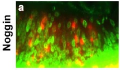
Noggin in Embryonic Mouse Cardiac Tissue. Noggin was detected in immersion fixed frozen sections of embryonic mouse cardiac tissue (11 d.p.c.) using 15 µg/mL Goat Anti-Mouse Noggin Antigen Affinity-purified Polyclonal Antibody (Catalog # AF719) overnight at 4 °C. Tissue was stained with the Anti-Goat HRP-DAB Cell & Tissue Staining Kit (brown; Catalog # CTS008) and counterstained with hematoxylin (blue). View our protocol for Chromogenic IHC Staining of Frozen Tissue Sections.
 View Larger
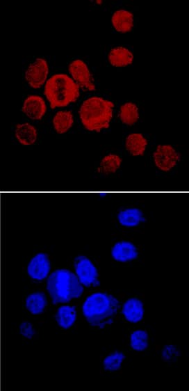
Noggin in PC‑3 Human Cell Line. Noggin was detected in immersion fixed PC-3 human prostate cancer cell line using Goat Anti-Mouse Noggin Antigen Affinity-purified Polyclonal Antibody (Catalog # AF719) at 10 µg/mL for 3 hours at room temperature. Cells were stained using the NorthernLights™ 557-conjugated Anti-Goat IgG Secondary Antibody (red, upper panel; Catalog # NL001) and counterstained with DAPI (blue, lower panel). Specific staining was localized to cytoplasm. View our protocol for Fluorescent ICC Staining of Cells on Coverslips.
 View Larger
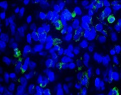
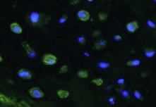
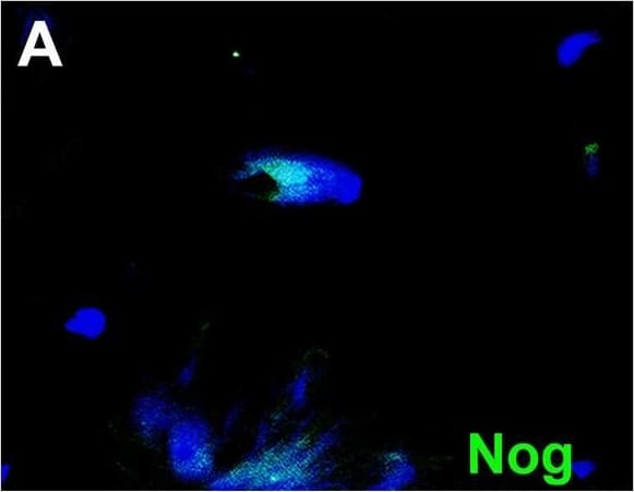
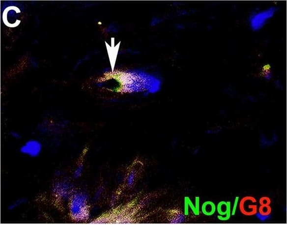
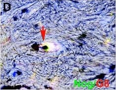
Detection of Human Noggin by Immunohistochemistry Myo/Nog cells contain ink in human tattooed skin.Tissue sections of tattooed skin were double labeled with the G8 mAb and antibodies to Noggin (Nog) (A-D), MyoD (F-I) or alpha -SMA (K-N). The colors of the secondary antibodies are indicated in the photographs. Nuclei were stained with Hoechst dye (blue). Images were produced with the epifluorescence (A-E, J and O) and confocal microscopes (F-I and K-N) with 60x lenses. Overlap of green and red fluorescence appears yellow in merged images (C, D, H, I, M and N). Fluorescent photomicrographs were merged with the corresponding DIC image to visualize the ink (black in D, I and N). Some double labeled cells appeared to contain ink (arrows in D, I and N). All ink laden G8+ cells were alpha -SMA+ (N). Smooth muscle cells of blood vessels also contained alpha -SMA (K). Minimal to no background fluorescence was visible after staining with the anti-goat (E), anti-IgM and anti-IgG (I), and anti-rabbit (O) secondary antibodies only. Bar = 28 μM in E and 5.6 μM in A-D and G-O. Image collected and cropped by CiteAb from the following open publication (https://pubmed.ncbi.nlm.nih.gov/32833999), licensed under a CC-BY license. Not internally tested by R&D Systems.
Preparation and Storage
- 12 months from date of receipt, -20 to -70 °C as supplied.
- 1 month, 2 to 8 °C under sterile conditions after reconstitution.
- 6 months, -20 to -70 °C under sterile conditions after reconstitution.
Background: Noggin
Noggin was originally cloned based on its dorsalizing activity in Xenopus embryos. Mammalian Noggins were subsequently identified and cloned from human, mouse and rat cDNA libraries. Mouse Noggin cDNA encodes a 232 amino acid (aa) residue precursor protein with 19 aa residue putative signal peptide that is cleaved to generate the 213 aa residue mature protein which is secreted as a homodimeric glycoprotein. Noggin is a highly conserved molecule. Mature mouse Noggin shares 99% and 83% aa sequence identity with human and Xenopus Noggin, respectively. Noggin has a complex pattern of expression during embryogenesis. In the adult, Noggin is expressed in the central nervous system and in several adult peripheral tissues such as lung, skeletal muscle and skin. Noggin has been shown to be a high-affinity BMP (bone morphogenetic protein) binding protein that antagonizes BMP bioactivities.
- Smith, W.C. and R.M. Harland (1992) Cell 70:829.
- Valenzuela, D.M. et al. (1995) J. Neurosci. 15:6077.
- Brunet, L.J. et al. (1998) Science 280:1455.
Product Datasheets
Citations for Mouse Noggin Antibody
R&D Systems personnel manually curate a database that contains references using R&D Systems products. The data collected includes not only links to publications in PubMed, but also provides information about sample types, species, and experimental conditions.
19
Citations: Showing 1 - 10
Filter your results:
Filter by:
-
Brain-specific angiogenesis inhibitor 1 is expressed in the Myo/Nog cell lineage
Authors: J Gerhart, J Bowers, L Gugerty, C Gerhart, M Martin, F Abdalla, A Bravo-Nuev, JT Sullivan, R Rimkunas, A Albertus, L Casta, L Getts, R Getts, M George-Wei
PLoS ONE, 2020-07-02;15(7):e0234792.
-
Taste papilla cell differentiation requires tongue mesenchyme via ALK3-BMP signaling to regulate the production of secretory proteins
Authors: M Ishan, Z Wang, P Zhao, Y Yao, S Stice, L Wells, Y Mishina, HX Liu
bioRxiv : the preprint server for biology, 2023-04-04;0(0):.
Species: Mouse
Sample Types: Whole Tissue
Applications: IHC -
Myo/Nog Cells Give Rise to Myofibroblasts During Epiretinal Membrane Formation in a Mouse Model of Proliferative Vitreoretinopathy
Authors: M Crispin, J Gerhart, A Heffer, M Martin, F Abdalla, A Bravo-Nuev, NJ Philp, AE Kuriyan, M George-Wei
Investigative Ophthalmology & Visual Science, 2023-02-01;64(2):1.
Species: Mouse
Sample Types: Whole Tissue
Applications: IHC -
Unveiling the transcriptomic landscape and the potential antagonist feedback mechanisms of TGF-beta superfamily signaling module in bone and osteoporosis
Authors: Ying-Wen Wang, Wen-Yu Lin, Fang-Ju Wu, Ching-Wei Luo
Cell Communication and Signaling
-
Acute Response and Neuroprotective Role of Myo/Nog Cells Assessed in a Rat Model of Focal Brain Injury
Authors: S Joseph-Pau, N Morrison, M Braccia, A Payne, L Gugerty, J Mostoller, P Lecker, EJ Tsai, J Kim, M Martin, R Brahmbhatt, G Gorski, J Gerhart, M George-Wei, J Stone, S Purushothu, A Bravo-Nuev
Frontiers in Neuroscience, 2021-12-07;15(0):780707.
Species: Rat
Sample Types: Whole Tissue
Applications: IHC -
A non-invasive method to generate induced pluripotent stem cells from primate urine
Authors: J Geuder, LE Wange, A Janjic, J Radmer, P Janssen, JW Bagnoli, S Müller, A Kaul, M Ohnuki, W Enard
Scientific Reports, 2021-02-10;11(1):3516.
Species: Human
Sample Types: Whole Cells
Applications: ICC -
Myo/Nog cells are nonprofessional phagocytes
Authors: J Gerhart, L Gugerty, P Lecker, F Abdalla, M Martin, O Gerhart, C Gerhart, K Johal, J Bernstein, J Spikes, K Mathers, A Bravo-Nuev, M George-Wei
PLoS ONE, 2020-08-24;15(8):e0235898.
Species: Human, Mouse
Sample Types: Whole Tissue
Applications: IHC -
Sphingosine 1-phosphate-regulated transcriptomes in heterogenous arterial and lymphatic endothelium of the aorta
Authors: Eric Engelbrecht, Michel V Levesque, Liqun He, Michael Vanlandewijck, Anja Nitzsche, Hira Niazi et al.
eLife
-
Rhabdomyosarcoma and Wilms tumors contain a subpopulation of noggin producing, myogenic cells immunoreactive for lens beaded filament proteins
Authors: J Gerhart, K Behling, M Paessler, L Milton, G Bramblett, D Garcia, M Pitts, R Hurtt, M Crawford, R Lackman, D Nguyen, J Infanti, P FitzGerald, M George-Wei
PLoS ONE, 2019-04-11;14(4):e0214758.
Species: Human
Sample Types: Whole Cells, Whole Tissue
Applications: ICC, IHC, IHC-P -
Noggin depletion in adipocytes promotes obesity in mice
Authors: AM Blázquez-M, M Jumabay, P Rajbhandar, T Sallam, Y Guo, J Yao, L Vergnes, K Reue, L Zhang, Y Yao, AM Fogelman, P Tontonoz, AJ Lusis, X Wu, KI Boström
Mol Metab, 2019-04-10;0(0):.
Species: Mouse
Sample Types: Whole Cells, Whole Tissue
Applications: Functional Assay, IHC -
Myo/Nog cells are present in the ciliary processes, on the zonule of Zinn and posterior capsule of the lens following cataract surgery
Authors: J Gerhart, C Withers, C Gerhart, L Werner, N Mamalis, A Bravo-Nuev, V Scheinfeld, P FitzGerald, R Getts, M George-Wei
Exp. Eye Res., 2018-03-17;171(0):101-105.
Species: Human, Mouse, Rabbit
Sample Types: Whole Tissue
Applications: IHC-P -
Antibody-Conjugated, DNA-Based Nanocarriers Intercalated with Doxorubicin Eliminate Myofibroblasts in Explants of Human Lens Tissue
Authors: Jacquelyn Gerhart, Marvin Greenbaum, Lou Casta, Anthony Clemente, Keith Mathers, Robert Getts et al.
Journal of Pharmacology and Experimental Therapeutics
-
Precise spatial restriction of BMP signaling is essential for articular cartilage differentiation
Authors: Ayan Ray, Pratik Narendra Pratap Singh, Michael L. Sohaskey, Richard M. Harland, Amitabha Bandyopadhyay
Development
-
Myo/Nog cells: targets for preventing the accumulation of skeletal muscle-like cells in the human lens.
Authors: Gerhart J, Greenbaum M, Scheinfeld V, Fitzgerald P, Crawford M, Bravo-Nuevo A, Pitts M, George-Weinstein M
PLoS ONE, 2014-04-15;9(4):e95262.
Species: Human
Sample Types: Whole Cells, Whole Tissue
Applications: ICC, IHC-Fr -
Joint TGF-beta type II receptor-expressing cells: ontogeny and characterization as joint progenitors.
Authors: Li T, Longobardi L, Myers T, Temple J, Chandler R, Ozkan H, Contaldo C, Spagnoli A
Stem Cells Dev, 2013-02-15;22(9):1342-59.
Species: Mouse
Sample Types: Whole Tissue
Applications: IHC -
Myo/Nog cell regulation of bone morphogenetic protein signaling in the blastocyst is essential for normal morphogenesis and striated muscle lineage specification
Authors: Jacquelyn Gerhart, Victoria L. Scheinfeld, Tara Milito, Jessica Pfautz, Christine Neely, Dakota Fisher-Vance et al.
Developmental Biology
-
BMP signalling permits population expansion by preventing premature myogenic differentiation in muscle satellite cells.
Authors: Ono Y, Calhabeu F, Morgan JE
Cell Death Differ., 2010-08-06;18(2):222-34.
Species: Mouse
Sample Types: Whole Cells
Applications: ICC -
Noggin producing, MyoD-positive cells are crucial for eye development.
Authors: Gerhart J, Pfautz J, Neely C, Elder J, DuPrey K, Menko AS, Knudsen K, George-Weinstein M
Dev. Biol., 2009-09-22;336(1):30-41.
Species: Chicken
Sample Types: Whole Tissue
Applications: IHC -
MyoD-positive epiblast cells regulate skeletal muscle differentiation in the embryo.
Authors: Gerhart J, Elder J, Neely C, Schure J, Kvist T, Knudsen K, George-Weinstein M
J. Cell Biol., 2006-10-23;175(2):283-92.
Species: Chicken
Sample Types: Whole Tissue
Applications: IHC-P
FAQs
No product specific FAQs exist for this product, however you may
View all Antibody FAQsReviews for Mouse Noggin Antibody
Average Rating: 5 (Based on 4 Reviews)
Have you used Mouse Noggin Antibody?
Submit a review and receive an Amazon gift card.
$25/€18/£15/$25CAN/¥75 Yuan/¥2500 Yen for a review with an image
$10/€7/£6/$10 CAD/¥70 Yuan/¥1110 Yen for a review without an image
Filter by:

