Mouse M-CSF Antibody Summary
Lys33-Glu262 (predicted)
Accession # P07141
Applications
Please Note: Optimal dilutions should be determined by each laboratory for each application. General Protocols are available in the Technical Information section on our website.
Scientific Data
 View Larger
Cell Proliferation Induced by M‑CSF and Neutralization by Mouse M‑CSF Antibody. Recombinant Mouse M-CSF (Catalog # 416-ML) stimulates proliferation in the M-NFS-60 mouse myelogenous luekemia lymphoblast cell line in a dose-dependent manner (orange line). Proliferation elicited by Recombinant Mouse M-CSF (10 ng/mL) is neutralized (green line) by increasing concentrations of Mouse M-CSF Monoclonal Antibody (Catalog # MAB4161). The ND50 is typically 3-12 µg/mL.
 View Larger
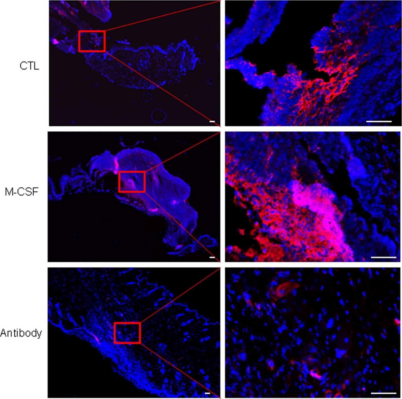
Detection of Mouse M-CSF by Immunocytochemistry/Immunofluorescence Injury-induced SSEA-1 positive cells were regulated by topical application of M-CSF or M-CSF neutralizing antibody.Skin wounds in mice were treated by either PBS control (top panels) or topical application of recombinant M-CSF protein (middle panels) or topical application of M-CSF neutralizing antibody for 6 days. This figure showed a representative skin section from 6 wounds in each group, stained with SSEA-1 (red color). DAPI (blue) was used for cell nucleus staining. Scale bars, 100 μm. Image collected and cropped by CiteAb from the following publication (https://www.nature.com/articles/srep28979), licensed under a CC-BY license. Not internally tested by R&D Systems.
 View Larger
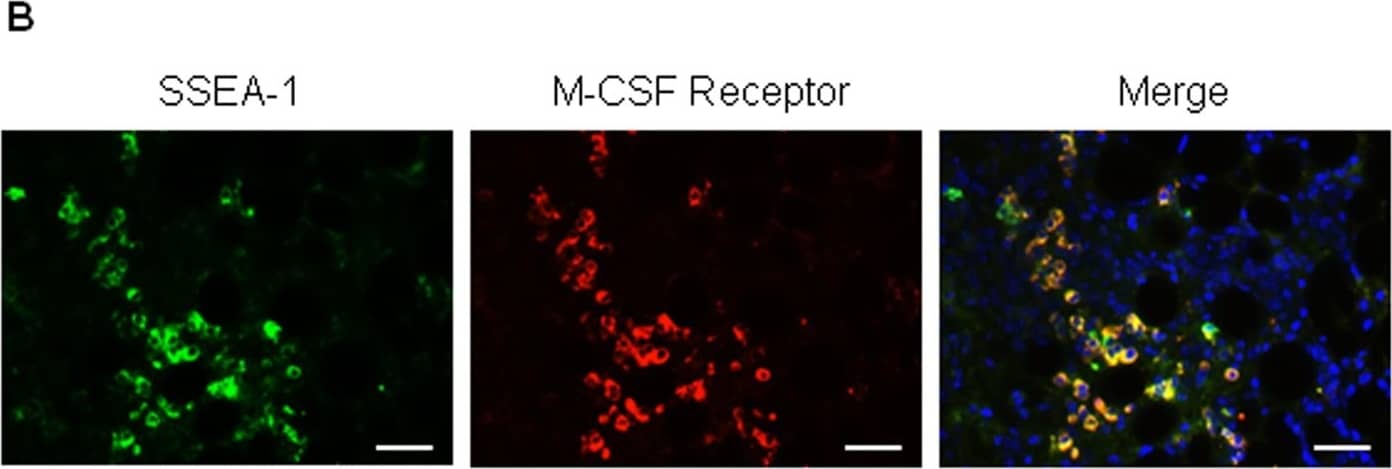
Detection of Mouse M-CSF by Immunocytochemistry/Immunofluorescence SSEA-1 positive cells are also SSEA-3 positive and express M-CSF receptor.(A) Injured skin sections were stained with SSEA-1 (red color) and SSEA-3 (green color) antibodies. IgG was used for a negative staining control. DAPI (blue) was used for cell nucleus staining. Scale bars, 50 μm. (B) Injured skin sections were stained with SSEA-1 (green color) and M-CSF receptor (red color) antibodies. DAPI (blue) was used for cell nucleus staining. Scale bars, 50 μm. Image collected and cropped by CiteAb from the following publication (https://www.nature.com/articles/srep28979), licensed under a CC-BY license. Not internally tested by R&D Systems.
Preparation and Storage
- 12 months from date of receipt, -20 to -70 °C as supplied.
- 1 month, 2 to 8 °C under sterile conditions after reconstitution.
- 6 months, -20 to -70 °C under sterile conditions after reconstitution.
Background: M-CSF
M-CSF, also known as CSF-1, is a four-alpha -helical-bundle cytokine that is the primary regulator of macrophage survival, proliferation and differentiation (1-3). M-CSF is also essential for the survival and proliferation of osteoclast progenitors (1, 4). M-CSF also primes and enhances macrophage killing of tumor cells and microorganisms, regulates the release of cytokines and other inflammatory modulators from macrophages, and stimulates pinocytosis (2, 3). M-CSF increases during pregnancy to support implantation and growth of the decidua and placenta (5). Sources of M-CSF include fibroblasts, activated macrophages, endometrial secretory epithelium, bone marrow stromal cells, and activated endothelial cells (1-5). The M-CSF receptor (c-fms) transduces its pleotropic effects and mediates its endocytosis. M-CSF mRNAs of various sizes occur (3-9). Full length mouse M-CSF transcripts encode a 520 amino acid (aa) type I transmembrane (TM) protein with a 462 aa extracellular region, a 21 aa TM domain, and a 37 aa cytoplasmic tail that forms a 140 kDa covalent dimer. Differential processing produces two proteolytically cleaved, secreted dimers. One is an N- and O-glycosylated 86 kDa dimer, while the other is modified by both glycosylation and chondroitin-sulfate proteoglycan (PG) to generate a 200 kDa subunit. Although PG-modified M-CSF can circulate, it may be immobilized by attachment to type V collagen (8). Shorter transcripts encode M-CSF that lacks cleavage and PG sites and produces an N-glycosylated 68 kDa TM dimer and a slowly produced 44 kDa secreted dimer (7). Although forms may vary in activity and half-life, all contain the N-terminal 150 aa portion that is necessary and sufficient for interaction with the M-CSF receptor (10, 11). The first 229 aa of mature mouse M-CSF shares 87%, 83%, 82%, and 81% aa identity with corresponding regions of rat, dog, cow, and human M-CSF, respectively (12, 13). Human M-CSF is active in the mouse, but mouse M-CSF is reported to be species-specific.
- Pixley, F.J. and E.R. Stanley (2004) Trends Cell Biol. 14:628.
- Chitu, V. and E.R. Stanley (2006) Curr. Opin. Immunol. 18:39.
- Fixe, P. and V. Praloran (1997) Eur. Cytokine Netw. 8:125.
- Ryan, G.R. et al. (2001) Blood 98:74.
- Makrigiannakis, A. et al. (2006) Trends Endocrinol. Metab. 17:178.
- Nandi, S. et al. (2006) Blood 107:786.
- Rettenmier, C.W. and M.F. Roussel (1988) Mol. Cell Biol. 8:5026.
- Suzu, S. et al. (1992) J. Biol. Chem. 267:16812.
- Manos, M.M. (1988) Mol. Cell. Biol. 8:5035.
- Koths, K. (1997) Mol. Reprod. Dev. 46:31.
- Jang, M-H. et al. (2006) J. Immunol. 177:4055.
- DeLamarter, J.F. et al. (1987) Nucleic Acids Res. 15:2389.
- Ladner, M.B. et al. (1988) Proc. Natl. Acad. Sci. USA 85:6706.
Product Datasheets
Citations for Mouse M-CSF Antibody
R&D Systems personnel manually curate a database that contains references using R&D Systems products. The data collected includes not only links to publications in PubMed, but also provides information about sample types, species, and experimental conditions.
7
Citations: Showing 1 - 7
Filter your results:
Filter by:
-
Enteric Glia Modulate Macrophage Phenotype and Visceral Sensitivity following Inflammation
Authors: V Grubiši?, JL McClain, DE Fried, I Grants, P Rajasekhar, E Csizmadia, OA Ajijola, RE Watson, DP Poole, SC Robson, FL Christofi, BD Gulbransen
Cell Rep, 2020-09-08;32(10):108100.
Species: Mouse
Sample Types: In situ
Applications: Activation -
Bone-derived Nestin-positive mesenchymal stem cells improve cardiac function via recruiting cardiac endothelial cells after myocardial infarction
Authors: D Lu, Y Liao, SH Zhu, QC Chen, DM Xie, JJ Liao, X Feng, MH Jiang, W He
Stem Cell Res Ther, 2019-04-27;10(1):127.
Species: N/A
Sample Types: Recombinant Protein
Applications: Western Blot -
Interleukin-18 Amplifies Macrophage Polarization and Morphological Alteration, Leading to Excessive Angiogenesis
Authors: T Kobori, S Hamasaki, A Kitaura, Y Yamazaki, T Nishinaka, A Niwa, S Nakao, H Wake, S Mori, T Yoshino, M Nishibori, H Takahashi
Front Immunol, 2018-03-06;9(0):334.
Species: Mouse
Sample Types: Cell Lysates, Whole Cells
Applications: Neutralization, Western Blot -
An inflammatory gene signature distinguishes neurofibroma Schwann cells and macrophages from cells in the normal peripheral nervous system
Authors: K Choi, K Komurov, JS Fletcher, E Jousma, JA Cancelas, J Wu, N Ratner
Sci Rep, 2017-03-03;7(0):43315.
Species: Mouse
Sample Types: Whole Cells
Applications: Neutralization -
CSF-1 receptor-mediated differentiation of a new type of monocytic cell with B cell-stimulating activity: its selective dependence on IL-34.
Authors: Yamane F, Nishikawa Y, Matsui K, Asakura M, Iwasaki E, Watanabe K, Tanimoto H, Sano H, Fujiwara Y, Stanley E, Kanayama N, Mabbott N, Magari M, Ohmori H
J Leukoc Biol, 2013-09-19;95(1):19-31.
Species: Mouse
Sample Types: Cell Lysates
Applications: Western Blot -
Colony-stimulating factor 1 receptor (CSF1R) signaling in injured neurons facilitates protection and survival.
Authors: Luo, Jian, Elwood, Fiona, Britschgi, Markus, Villeda, Saul, Zhang, Hui, Ding, Zhaoqing, Zhu, Liyin, Alabsi, Haitham, Getachew, Ruth, Narasimhan, Ramya, Wabl, Rafael, Fainberg, Nina, James, Michelle, Wong, Gordon, Relton, Jane, Gambhir, Sanjiv S, Pollard, Jeffrey, Wyss-Coray, Tony
J Exp Med, 2013-01-07;210(1):157-72.
Species: Mouse
Sample Types: Whole Tissue
Applications: IHC-Fr -
Osteoblasts support B-lymphocyte commitment and differentiation from hematopoietic stem cells.
Authors: Zhu J, Garrett R, Jung Y, Zhang Y, Kim N, Wang J, Joe GJ, Hexner E, Choi Y, Taichman RS, Emerson SG
Blood, 2007-01-16;109(9):3706-12.
Species: Mouse
Sample Types: Whole Cells
Applications: Neutralization
FAQs
No product specific FAQs exist for this product, however you may
View all Antibody FAQsReviews for Mouse M-CSF Antibody
There are currently no reviews for this product. Be the first to review Mouse M-CSF Antibody and earn rewards!
Have you used Mouse M-CSF Antibody?
Submit a review and receive an Amazon gift card.
$25/€18/£15/$25CAN/¥75 Yuan/¥2500 Yen for a review with an image
$10/€7/£6/$10 CAD/¥70 Yuan/¥1110 Yen for a review without an image


