Human TIM-3 Antibody Summary
Met1-Arg200
Accession # Q8TDQ0
Applications
Please Note: Optimal dilutions should be determined by each laboratory for each application. General Protocols are available in the Technical Information section on our website.
Scientific Data
 View Larger
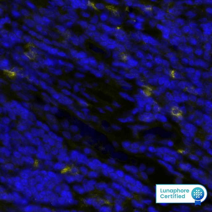
Detection of TIM-3 in Human Tonsil via seqIF™ staining on COMET™ TIM-3 was detected in immersion fixed paraffin-embedded sections of human Tonsil using Rabbit Anti-Human TIM-3, Monoclonal Antibody (Catalog #MAB11672) at 15ug/mL at 37 ° Celsius for 4 minutes. Before incubation with the primary antibody, tissue underwent an all-in-one dewaxing and antigen retrieval preprocessing using PreTreatment Module (PT Module) and Dewax and HIER Buffer H (pH 9; Epredia Catalog # TA-999-DHBH). Tissue was stained using the Alexa Fluor™ Plus 647 Goat anti-Rabbit IgG Secondary Antibody at 1:200 at 37 ° Celsius for 2 minutes. (Yellow; Lunaphore Catalog # DR647RB) and counterstained with DAPI (blue; Lunaphore Catalog # DR100). Specific staining was localized to the membrane. Protocol available in COMET™ Panel Builder.
 View Larger
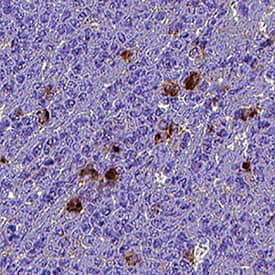
Detection of TIM‑3 in Human Tonsil. TIM‑3 was detected in immersion fixed paraffin-embedded sections of human tonsil using Rabbit Anti-Human TIM‑3 Monoclonal Antibody (Catalog # MAB11672) at 3 µg/ml for 1 hour at room temperature followed by incubation with the Anti-Rabbit IgG VisUCyte™ HRP Polymer Antibody (Catalog # VC003) or the HRP-conjugated Anti-Rabbit IgG Secondary Antibody (Catalog # HAF008). Before incubation with the primary antibody, tissue was subjected to heat-induced epitope retrieval using VisUCyte Antigen Retrieval Reagent-Basic (Catalog # VCTS021). Tissue was stained using DAB (brown) and counterstained with hematoxylin (blue). Specific staining was localized to the cell membrane. View our protocol for IHC Staining with VisUCyte HRP Polymer Detection Reagents.
Preparation and Storage
- 12 months from date of receipt, -20 to -70 °C as supplied.
- 1 month, 2 to 8 °C under sterile conditions after reconstitution.
- 6 months, -20 to -70 °C under sterile conditions after reconstitution.
Background: TIM-3
TIM-3 (T cell immunoglobulin and mucin domain-3) is a 60 kDa member of the TIM family of immune regulating molecules. TIMs are type I transmembrane glycoproteins with one Ig-like V-type domain and a Ser/Thr-rich mucin stalk (1-3). There are three TIM genes in human and eight in mouse. Mature human TIM-3 consists of a 181 amino acid (aa) extracellular domain (ECD), a 21 aa transmembrane segment, and a 78 aa cytoplasmic tail (4). An alternately spliced isoform is truncated following a short substitution after the Ig-like domain. Within the ECD, human TIM-3 shares 58% aa sequence identity with mouse and rat TIM-3. TIM-3 is expressed on the surface of effector T cells (CD4+ Th1 and CD8+ Tc1) but not on helper T cells (CD4+ Th2 and CD8+ Tc2) (4, 5). NK cells appear to transcribe the highest amounts of Tim-3 among lymphocytes, and when Tim-3 was cross-linked with antibodies it suppressed NK cell-mediated cytotoxicity (6). In chronic inflammation, autoimmune disorders, and some cancers, TIM-3 is upregulated on several other hematopoietic cell types. It also occurs on hippocampal neurons (7-10). The Ig domain of TIM-3 interacts with a ligand on resting but not activated Th1 and Th2 cells (5, 11). The glycosylated Ig domain of TIM-3 binds cell-associated galectin-9. This induces TIM-3 Tyr phosphorylation and pro-apoptotic signaling (8, 12). TIM-3 functions as a negative regulator of Th1 cell activity. Its blockade results in increased IFN-gamma production, Th1 cell proliferation and cytotoxicity (5, 10, 11), and regulatory T cell development (5). TIM-3 inhibits the antitumor efficacy of DNA vaccines and chemotherapy
by binding to the damage-associated molecular pattern molecule, HMGB1 (13).
- Anderson, A.C. and D.E. Anderson (2006) Curr. Opin. Immunol. 18:665.
- Mariat, C. et al. (2005) Phil. Trans. R. Soc. B. 360:1681.
- Meyers, J.H. et al. (2005) Trends Mol. Med. 11:362.
- Monney, L. et al. (2002) Nature 415:536.
- Sanchez-Fueyo, A. et al. (2003) Nat. Immunol. 4:1093.
- Ndhlovu, L. et al. (2012) Blood 119:3734.
- Wiener, Z. et al. (2007) J. Invest. Dermatol. 127:906.
- van de Weyer, P.S. et al. (2006) Biochem. Biophys. Res. Commun. 351:571.
- Gielen, A.W. et al. (2005) J. Neuroimmunol. 164:93.
- Oikawa, T. et al. (2006) J. Immunol. 177:4281.
- Sabatos, C.A. et al. (2003) Nat. Immunol. 4:1102.
- Zhu, C. et al. (2005) Nat. Immunol. 6:1245.
- Chiba, S. et al. (2012) Nat. Immunol. 13:832.
Product Datasheets
FAQs
No product specific FAQs exist for this product, however you may
View all Antibody FAQsReviews for Human TIM-3 Antibody
There are currently no reviews for this product. Be the first to review Human TIM-3 Antibody and earn rewards!
Have you used Human TIM-3 Antibody?
Submit a review and receive an Amazon gift card.
$25/€18/£15/$25CAN/¥75 Yuan/¥2500 Yen for a review with an image
$10/€7/£6/$10 CAD/¥70 Yuan/¥1110 Yen for a review without an image
