Human RIPK3/RIP3 Antibody Summary
Met1-Arg218
Accession # Q9Y572
Applications
Please Note: Optimal dilutions should be determined by each laboratory for each application. General Protocols are available in the Technical Information section on our website.
Scientific Data
 View Larger
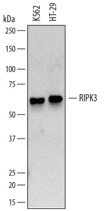
Detection of Human RIPK3/RIP3 by Western Blot. Western blot shows lysates of K562 human chronic myelogenous leukemia cell line and HT-29 human colon adenocarcinoma cell line. PVDF membrane was probed with 1 µg/mL of Mouse Anti-Human RIPK3/RIP3 Monoclonal Antibody (Catalog # MAB7604) followed by HRP-conjugated Anti-Mouse IgG Secondary Antibody (Catalog # HAF007). A specific band was detected for RIPK3/RIP3 at approximately 60 kDa (as indicated). This experiment was conducted under reducing conditions and using Immunoblot Buffer Group 1.
 View Larger
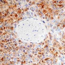
RIPK3/RIP3 in Human Spleen. RIPK3/RIP3 was detected in immersion fixed paraffin-embedded sections of human spleen using Mouse Anti-Human RIPK3/RIP3 Monoclonal Antibody (Catalog # MAB7604) at 15 µg/mL overnight at 4 °C. Tissue was stained using the Anti-Mouse HRP-DAB Cell & Tissue Staining Kit (brown; Catalog # CTS002) and counterstained with hematoxylin (blue). Specific staining was localized to the cytoplasm of splenocytes. View our protocol for Chromogenic IHC Staining of Paraffin-embedded Tissue Sections.
Preparation and Storage
- 12 months from date of receipt, -20 to -70 °C as supplied.
- 1 month, 2 to 8 °C under sterile conditions after reconstitution.
- 6 months, -20 to -70 °C under sterile conditions after reconstitution.
Background: RIPK3/RIP3
RIP3 (Receptor-Interacting Protein 3), also known as RIPK3, is a 518 amino acid (aa), 60 kDa phosphoprotein that contains an N-terminal protein kinase domain and a C-terminal interaction motif with which it binds RIP1. It converts cell response to TNF-a from apoptosis (RIP1 via Fas) to programmed necrosis (RIP1/RIP3) by inducing reactive oxygen species production. Human RIP3 (aa 1-218) shares approximately 74% aa sequence identity with mouse and rat RIP3. Alternately spliced human b (252 aa) and g (231 aa) isoforms diverge after aa 219 and 221, respectively, and are found to antagonize RIP3.
Product Datasheets
Citations for Human RIPK3/RIP3 Antibody
R&D Systems personnel manually curate a database that contains references using R&D Systems products. The data collected includes not only links to publications in PubMed, but also provides information about sample types, species, and experimental conditions.
4
Citations: Showing 1 - 4
Filter your results:
Filter by:
-
Necroptosis contributes to the intestinal toxicity of deoxynivalenol and is mediated by methyltransferase SETDB1
Authors: Zhou, B;Xiao, K;Guo, J;Xu, Q;Xu, Q;Lv, Q;Zhu, H;Zhao, J;Liu, Y;
Journal of hazardous materials
Species: Porcine
Sample Types: Cell Lysates, Tissue Homogenates
Applications: Western Blot -
Acyl-Coenzyme A Synthetase Long-Chain Family Member 4 Is Involved in Viral Replication Organelle Formation and Facilitates Virus Replication via Ferroptosis
Authors: YA Kung, HJ Chiang, ML Li, YN Gong, HP Chiu, CT Hung, PN Huang, SY Huang, PY Wang, TA Hsu, G Brewer, SR Shih
MBio, 2022-01-18;0(0):e0271721.
Species: Human
Sample Types: Cell Lysates
Applications: Western Blot -
ABT?737, a Bcl?2 family inhibitor, has a synergistic effect with apoptosis by inducing urothelial carcinoma cell necroptosis
Authors: R Cheng, X Liu, Z Wang, K Tang
Molecular Medicine Reports, 2021-03-31;23(6):.
Species: Human
Sample Types: Cell Lysates
Applications: Co-Immunoprecipitation, Western Blot -
Prognostic Significance of CHIP and RIPK3 in Non-Small Cell Lung Cancer
Authors: J Kim, JY Chung, YS Park, SJ Jang, HR Kim, CM Choi, JS Song
Cancers (Basel), 2020-06-08;12(6):.
Species: Human
Sample Types: Whole Tissue
Applications: IHC
FAQs
No product specific FAQs exist for this product, however you may
View all Antibody FAQsReviews for Human RIPK3/RIP3 Antibody
Average Rating: 5 (Based on 1 Review)
Have you used Human RIPK3/RIP3 Antibody?
Submit a review and receive an Amazon gift card.
$25/€18/£15/$25CAN/¥75 Yuan/¥2500 Yen for a review with an image
$10/€7/£6/$10 CAD/¥70 Yuan/¥1110 Yen for a review without an image
Filter by:

