Human Phospho-STAT3 (Y705) Antibody Summary
Applications
Please Note: Optimal dilutions should be determined by each laboratory for each application. General Protocols are available in the Technical Information section on our website.
Scientific Data
 View Larger
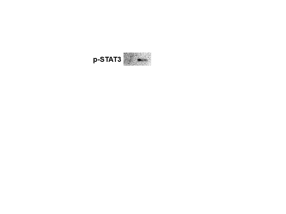
Detection of Human Phospho-STAT3 (Y705) by Western Blot. Western blot shows lysates of Daudi human Burkitt's lymphoma cell line and HepG2 human hepatocellular carcinoma cell line untreated (-) or treated (+) with 500 U/mL Recombinant Human IFN-aA (Catalog # 11100-1) for 20 minutes or 50 µg/mL Recombinant Human IL-22 (Catalog # 782-IL) for 15 minutes. PVDF membrane was probed with 0.5 µg/mL of Rabbit Anti-Human Phospho-STAT3 (Y705) Antigen Affinity-purified Polyclonal Antibody (Catalog # AF4607) followed by HRP-conjugated Anti-Rabbit IgG Secondary Antibody (Catalog # HAF008). A specific band was detected for STAT3 at approximately 95 kDa (as indicated). This experiment was conducted under reducing conditions and using Immunoblot Buffer Group 1.
 View Larger
Detection of Phospho-STAT3 (Y705) in IFN-alpha-treated Human Daudi Cell Line by Flow Cytometry. Daudi human Burkitt's lymphoma cell line was unstimulated (light orange filled histogram) or treated with 500 U/mL rhIFN-alpha for 20 minutes (dark orange filled histogram) was stained with Human Phospho-STAT3 (Y705) Antigen Affinity-purified Polyclonal Antibody (Catalog # AF4607) or control antibody (Catalog # AB-105-C, open histogram), followed by Phycoerythrin-conjugated Anti-Rabbit IgG Secondary Antibody (Catalog # F0110). To facilitate intracellular staining, cells were fixed with paraformaldehyde and permeabilized with methanol.
 View Larger
STAT3 in Daudi Human Burkitt's Lymphoma Cells. Phospho-STAT3 was detected in immersion fixed IFN-alpha treated Daudi human Burkitt's lymphoma cell line using 10 µg/mL Human Phospho-STAT3 (Y705) Antigen Affinity-purified Polyclonal Antibody (Catalog # AF4607) for 3 hours at room temperature. Cells were stained with the NorthernLights™ 557-conjugated Anti-Rabbit IgG Secondary Antibody (red; Catalog # NL004) and counterstained with DAPI (blue). View our protocol for Fluorescent ICC Staining of Cells on Coverslips.
 View Larger
Detection of Human STAT3 by Simple WesternTM. Simple Western shows lysates of Daudi human Burkitt's lymphoma cell line untreated (-) or treated (+) with 50 µg/mL Recombinant Human IL‑22 (Catalog # 782-IL) for 15 minutes, loaded at 0.2 mg/ml. A specific band was detected for STAT3 at approximately 91 kDa (as indicated) using 10 µg/mL of Rabbit Anti-Human Phospho-STAT3 (Y705) Antigen Affinity-purified Polyclonal Antibody (Catalog # AF4607). This experiment was conducted under reducing conditions and using the 12-230kDa separation system.
 View Larger
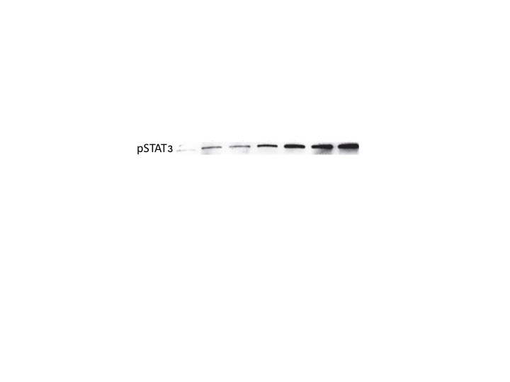
Detection of Human STAT3 by Western Blot IL-22BPi1 does not interact with IL-22 or IL-22BPi2. (A) A549 cells were exposed for 30 minutes to dilutions of IL-22-containing culture medium (CM) previously produced by IL22-transfected HEK293 cells. A549 cell lysates (CL) were immunoblotted for pSTAT3 and tubulin as loading control. The relative densitometry of pSTAT3 normalized to that of tubulin is also represented. (B) A549 cells were treated with the optimum IL-22 dilution from A (1/512) for different periods of time, lysed and immunoblotted for pSTAT3. An unspecific protein band (u.p.) was used as loading control. (C) IL-22BP concentration in conditioned medium (CM) of transfected HEK293 cells was measured by ELISA, and the indicated amounts in nanograms (ng) of IL-22BPi1 or IL-22BPi2 were pre-incubated for 1 h at 37°C with the selected IL-22 concentration from (A). A549 were exposed to the pre-incubated combinations for 20 min. An excess of IL-22BPi2 was used as phosphorylation blocking control (lane 3). Cells were lysed and immunoblotted for pSTAT3 and actin as loading control. (D) HeLa cells were co-transfected with the indicated expression plasmids, 24 h later cells were lysed and the CM was subjected to acetone precipitation (AP), and proteins were immunoblotted for FLAG. Intracellular IL-22BPi1 is indicated with dark purple arrows, intracellular and secreted IL-22BPi2 is indicated with green arrows, and co-expressed IL-22 and IL-17 are also indicated. (E) Conditioned media (CM) from 3 independent experiments, in which expression vectors for the three IL-22BP isoforms were individually transfected into HEK293 cells together with either IL-17 or IL-22 expression vectors or an empty vector control (EV), were analyzed 24 h after transfection by ELISA for IL-22BP (mean ± SEM; n = 3; **p < 0.01 by unpaired t-test). (F) IL-22BPi1 does not interact with IL-22BPi2. IL-22BPi1-MF expression plasmid containing Myc and FLAG tags was co-transfected with an inducible pTRE3G-based vector expressing IL-22BPi2 with only a FLAG tag. After 24 h, cells were induced for IL-22BPi2 production by adding Tet-Express activator to the medium for a further 24 h. Cells were lysed and immunoprecipitated with anti-Myc agarose, the flow-through fractions were then further subjected to FLAG immunoprecipitation. CL and eluted fractions were immunoblotted for FLAG. Image collected and cropped by CiteAb from the following publication (https://pubmed.ncbi.nlm.nih.gov/30619294), licensed under a CC-BY license. Not internally tested by R&D Systems.
 View Larger
Detection of Human Human Phospho-STAT3 (Y705) Antibody by Western Blot N-EV and H-EV treatment promote macrophage M2 polarization by delivering miR-21-5p that targets PTEN. a, western blot analysis of PTEN protein expression level in induced macrophages. H/i-miR-EV, monocytes were induced with the presence of EV secreted by miR-21-5p-inhibited, hypoxia pre-challenged MSCs; H-EV + i-miR, monocytes were transfected with miR-21-5p inhibitor-expressing vector before induction with the presence of H-EV. Macrophages induced without MSC-EV were used as negative control (NC). b, c, flow cytometry determining the percentage of CD163+CD206+ cells among total CD68+ cells after induction. N-EV + O/E PTEN or H-EV + O/E PTEN, monocytes were transfected with PTEN overexpressing vector before N-EV or H-EV treatment, respectively. d–f, western blot detecting Akt and STAT3 protein expression as well as their activating phosphorylation (p-Ser473 for Akt and p-tyr705 for STAT3) in macrophages after induction. g–i, ELISA evaluating IL-10, TGF-beta and VEGF-alpha in macrophage culture medium after induction. Macrophages induced with the presence of N-EV were used as negative control in b–i. Tukey’s test was used for statistical analysis. *, p < 0.05; **, p < 0.01; ***, p < 0.001; ****, p < 0.0001 Image collected and cropped by CiteAb from the following publication (https://pubmed.ncbi.nlm.nih.gov/30736829), licensed under a CC-BY license. Not internally tested by R&D Systems.
Preparation and Storage
- 12 months from date of receipt, -20 to -70 °C as supplied.
- 1 month, 2 to 8 °C under sterile conditions after reconstitution.
- 6 months, -20 to -70 °C under sterile conditions after reconstitution.
Background: STAT3
Signal Transducer and Activator of Transcription (STAT) proteins are transcription factors activated in response to cytokine, growth factor, or hormone receptor signaling. Janus kinases (JAKs) phosphorylate STAT proteins and induce dimerization. Homo- or heterodimers translocate to the nucleus where they bind to DNA and activate transcription.
Product Datasheets
Citations for Human Phospho-STAT3 (Y705) Antibody
R&D Systems personnel manually curate a database that contains references using R&D Systems products. The data collected includes not only links to publications in PubMed, but also provides information about sample types, species, and experimental conditions.
8
Citations: Showing 1 - 8
Filter your results:
Filter by:
-
Thymoquinone Inhibits JAK/STAT and PI3K/Akt/ mTOR Signaling Pathways in MV4-11 and K562 Myeloid Leukemia Cells
Authors: Futoon Abedrabbu Al-Rawashde, Abdullah Saleh Al-wajeeh, Mansoureh Nazari Vishkaei, Hanan Kamel M. Saad, Muhammad Farid Johan, Wan Rohani Wan Taib et al.
Pharmaceuticals (Basel)
-
Interleukin-6 Signaling in Triple Negative Breast Cancer Cells Elicits the Annexin A1/Formyl Peptide Receptor 1 Axis and Affects the Tumor Microenvironment
Authors: L Vecchi, STS Mota, MAP Zóia, IC Martins, JB de Souza, TG Santos, AO Beserra, VP de Andrade, LR Goulart, TG Araújo
Cells, 2022-05-20;11(10):.
Species: Human
Sample Types: Whole Cells
Applications: Flow Cytometry -
LIMD2 Regulates Key Steps of Metastasis Cascade in Papillary Thyroid Cancer Cells via MAPK Crosstalk
Authors: RP Araldi, TC de Melo, D Levy, DM de Souza, B Maurício, GA Colozza-Ga, SP Bydlowski, H Peng, FJ Rauscher, JM Cerutti
Cells, 2020-11-23;9(11):.
Species: Human
Sample Types: Whole Cells
Applications: Flow Cytometry -
Circular RNA circHIPK3 modulates autophagy via MIR124-3p-STAT3-PRKAA/AMPK? signaling in STK11 mutant lung cancer
Authors: X Chen, R Mao, W Su, X Yang, Q Geng, C Guo, Z Wang, J Wang, LA Kresty, DG Beer, AC Chang, G Chen
Autophagy, 2019-06-28;0(0):1-13.
Species: Human
Sample Types: Cell Lysates
Applications: Western Blot -
Extracellular vesicles secreted by hypoxia pre-challenged mesenchymal stem cells promote non-small cell lung cancer cell growth and mobility as well as macrophage M2 polarization via miR-21-5p delivery
Authors: W Ren, J Hou, C Yang, H Wang, S Wu, Y Wu, X Zhao, C Lu
J. Exp. Clin. Cancer Res., 2019-02-08;38(1):62.
Species: Human
Sample Types: Cell Lysates
Applications: Western Blot -
IL-6 cytoprotection in hyperoxic acute lung injury occurs via suppressor of cytokine signaling-1-induced apoptosis signal-regulating kinase-1 degradation.
Authors: Kolliputi N, Waxman AB
Am. J. Respir. Cell Mol. Biol., 2008-09-05;40(3):314-24.
Species: Human
Sample Types: Cell Lysates
Applications: Western Blot -
Long Interleukin-22 Binding Protein Isoform-1 Is an Intracellular Activator of the Unfolded Protein Response
Authors: Paloma Gómez-Fernández, Andoni Urtasun, Adrienne W. Paton, James C. Paton, Francisco Borrego, Devin Dersh et al.
Frontiers in Immunology
-
Leptin promotes a proinflammatory lipid profile and induces inflammatory pathways in human SZ95 sebocytes
Authors: D. Törőcsik, D. Kovács, E. Camera, M. Lovászi, K. Cseri, G.G. Nagy et al.
British Journal of Dermatology
FAQs
No product specific FAQs exist for this product, however you may
View all Antibody FAQsReviews for Human Phospho-STAT3 (Y705) Antibody
Average Rating: 5 (Based on 2 Reviews)
Have you used Human Phospho-STAT3 (Y705) Antibody?
Submit a review and receive an Amazon gift card.
$25/€18/£15/$25CAN/¥75 Yuan/¥2500 Yen for a review with an image
$10/€7/£6/$10 CAD/¥70 Yuan/¥1110 Yen for a review without an image
Filter by:


