Human PDGF R beta Antibody Summary
*Small pack size (-SP) is supplied either lyophilized or as a 0.2 µm filtered solution in PBS.
Applications
Please Note: Optimal dilutions should be determined by each laboratory for each application. General Protocols are available in the Technical Information section on our website.
Scientific Data
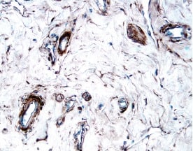 View Larger
PDGF R beta in Human Breast Cancer Tissue. PDGF R beta was detected in immersion fixed paraffin-embedded sections of human breast cancer tissue using 15 µg/mL Goat Anti-Human PDGF R beta Antigen Affinity-purified Polyclonal Antibody (Catalog # AF385) overnight at 4 °C. Tissue was stained with the Anti-Goat HRP-DAB Cell & Tissue Staining Kit (brown; CTS008) and counterstained with hematoxylin (blue). View our protocol for Chromogenic IHC Staining of Paraffin-embedded Tissue Sections.
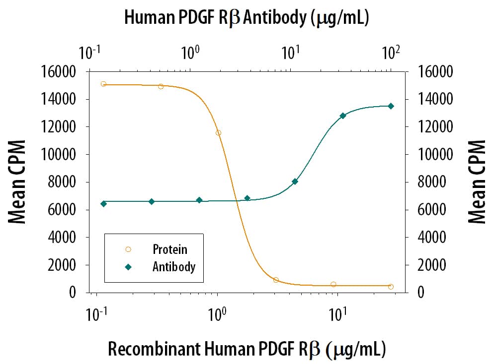 View Larger
PDGF R beta Inhibition of PDGF-BB-dependent Cell Proliferation and Neutralization by Human PDGF R beta Antibody. Recombinant Human PDGF R beta Fc Chimera (385-PR) inhibits Recombinant Human PDGF-BB (220-BB) induced proliferation in the NR6R-3T3 mouse fibroblast cell line in a dose-dependent manner (orange line). Inhibition of Recombinant Human PDGF-BB (4 ng/mL) activity elicited by Recombinant Human PDGF R beta Fc Chimera (2 µg/mL) is neutralized (green line) by increasing concentrations of Goat Anti-Human PDGF R beta Antigen Affinity-purified Polyclonal Antibody (Catalog # AF385). The ND50 is typically 10-40 µg/mL.
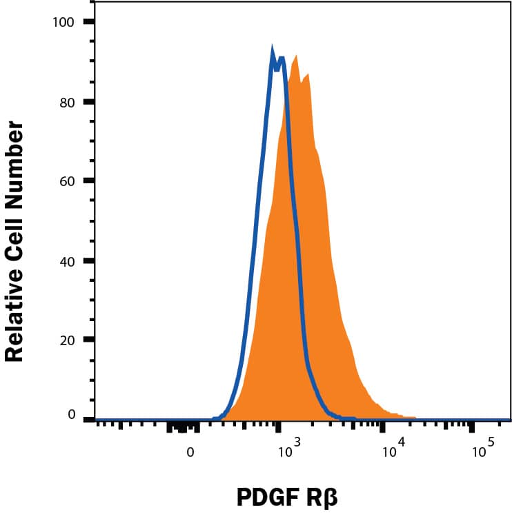 View Larger
Detection of PDGF R beta in U87MG cells by Flow Cytometry. U87MG cells were stained with Goat Anti-Human PDGF R beta Antigen Affinity-purified Polyclonal Antibody (Catalog # AF385, filled histogram) or isotype control antibody (Catalog # AB-108-C, open histogram), followed by Phycoerythrin-conjugated Anti-Goat IgG Secondary Antibody (Catalog # F0107). View our protocol for Staining Membrane-associated Proteins.
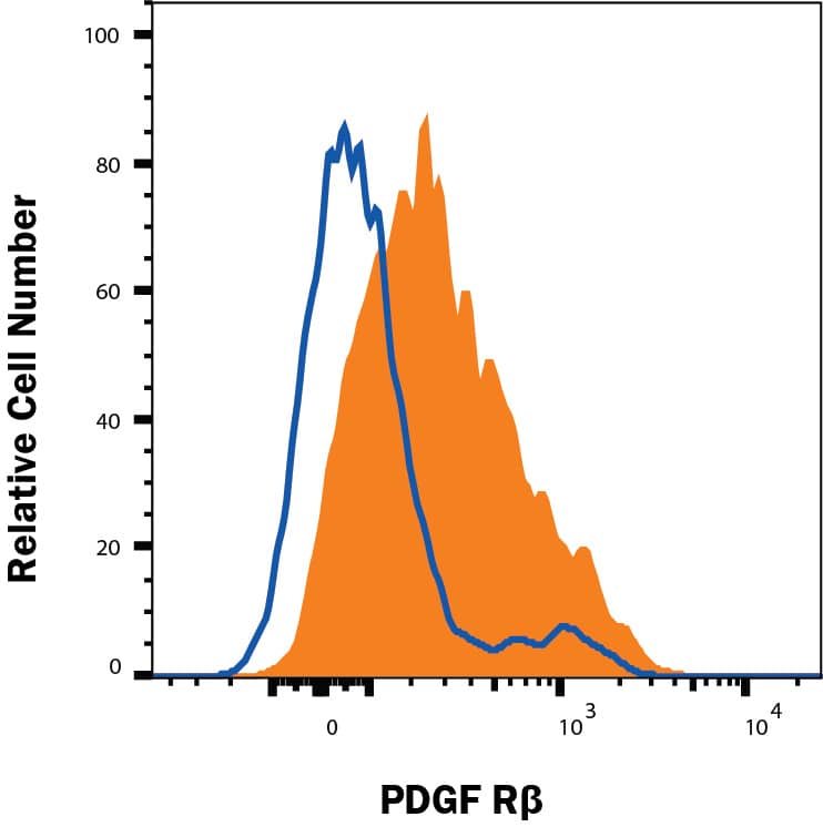 View Larger
Detection of PDGF R beta in U118MG cells by Flow Cytometry. U118MG cells were stained with Goat Anti-Human PDGF R beta Antigen Affinity-purified Polyclonal Antibody (Catalog # AF385, filled histogram) or isotype control antibody (Catalog # AB-108-C, open histogram), followed by Phycoerythrin-conjugated Anti-Goat IgG Secondary Antibody (Catalog # F0107). View our protocol for Staining Membrane-associated Proteins.
 View Larger
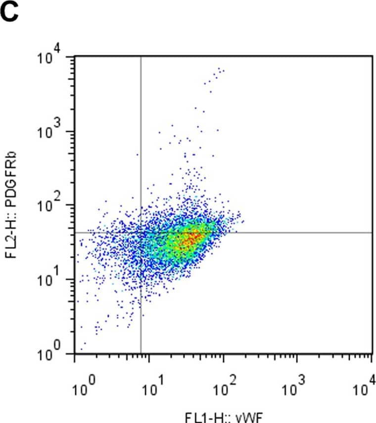
Detection of Human PDGF R beta by Flow Cytometry Co-expression of EC marker and PDGFR beta after EPC-CM incubation.Co-expression of vWF and PDGFR beta was measured using dual color FACS analysis. HUVEC were kept in control medium containing only 1% FCS or EPC-CM for 24 h before the measurement. HUVEC incubated in control medium showed only a fractional amount of vWF+/PDGFR beta + cells (A). After EPC-CM exposure the proportion of vWF+/PDGFR beta + double positive population was significantly increased, suggesting a strong phenotype shift of endothelial cells towards PDGFR beta + (B). The addition of neutralizing antibody AF385 did not block the upreguation of the PDGFR beta by EPC-CM stimulation (C). However, such enhanced PDGFR beta expression could not be evoked by solely adding 100 ng/ml (D) or 100 pg/ml (E) rhPDGF-BB to the control medium. *, P<0.01 compared to controls. Image collected and cropped by CiteAb from the following publication (https://dx.plos.org/10.1371/journal.pone.0014107), licensed under a CC-BY license. Not internally tested by R&D Systems.
 View Larger
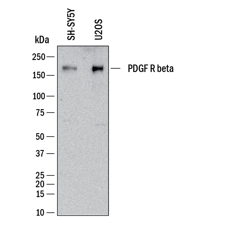
Detection of Human PDGF R beta by Western Blot. Western Blot shows lysates of SH‑SY5Y human neuroblastoma cell line and U2OS human osteosarcoma cell line. PVDF membrane was probed with 2 µg/ml of Goat Anti-Human PDGF R beta Antigen Affinity-purified Polyclonal Antibody (Catalog # AF385) followed by HRP-conjugated Anti-Goat IgG Secondary Antibody (Catalog # HAF017). A specific band was detected for PDGF R beta at approximately 190 kDa (as indicated). This experiment was conducted under reducing conditions and using Western Blot Buffer Group 1.
 View Larger
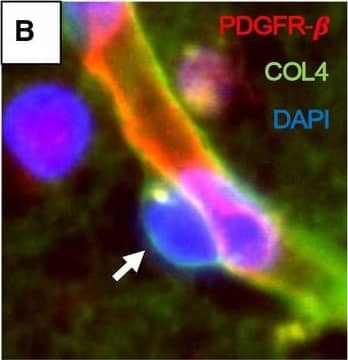
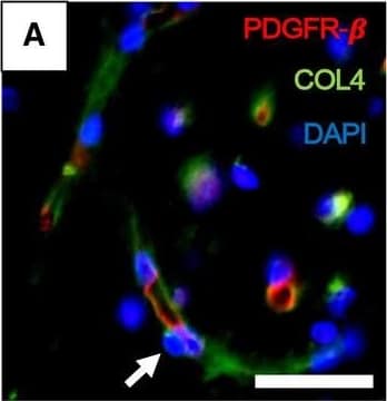
Detection of Mouse PDGF R beta by Immunohistochemistry Pericyte soma on WM capillaries demonstrated by immunofluorescence. A–F. PDGFR‐ beta (red) and COL4 (green) immunofluorescence staining with nuclei (DAPI) (blue). A,B. Low‐ and high‐power images showing capillary segments (arrowheads) with overlapping PDGFR‐ beta (red), COL4 (green) and DAPI (blue). C. Same vessel segment as B with PDGFR‐ beta (red) and COL4 (green); D. COL4 (green) and DAPI (blue); E. PDGFR‐ beta (red) and DAPI (blue). F. Another capillary segment with PDGFR‐ beta (red), COL4 (green) and DAPI (blue) clearly showing pericyte cell body. G–J. Images taken by a confocal microscope showing pericyte cell bodies (arrows) with nuclear stain (DAPI). Capillaries and pericyte processes are revealed by COL4 and PDGFR‐ beta (red) immunoreactivities. Magnification bars: A = 50 µm, F, J = 10 µm. Image collected and cropped by CiteAb from the following open publication (https://pubmed.ncbi.nlm.nih.gov/32705757), licensed under a CC-BY license. Not internally tested by R&D Systems.
 View Larger
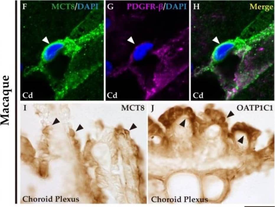
Detection of Rhesus Macaque PDGF R beta by Immunohistochemistry Expression of MCT8 and OATP1C1 in blood vessels and brain barriers in the human and macaque basal ganglia and adjacent choroid plexus. (A) Representative brightfield photomicrograph shows immunostaining for MCT8 in the human putamen. Note that an MCT8 immunopositive signal is observed along the capillary wall (red arrowhead), fibers (green arrowhead), and “bump-on-a-log” morphology pericytelike cells (white arrowhead). (B–H) Representative confocal microscope compositions from multiple-stained sections for MCT8 (green), the endothelial marker UEA-I (red), and the vascular and pericyte biomarker PDGFR-beta (purple) in human and macaque caudate nucleus. Merged image (E,H) shows the colocalization of all signals. (B–E) Coexpression of MCT8, UEA-I, and PDGFR-beta is observed in a vessel, while coexpression of MCT8 and PDGFR-beta but not UEA-I is observed in a capillary-associated pericyte (white arrowheads) in humans. (F–H) Coexpression of MCT8 and PDGFR-beta in a vessel and pericytelike cells (white arrowheads) in macaques. Counterstaining with DAPI (blue) shows nuclei of all cells. (I,J) Representative brightfield photomicrographs show immunostaining for MCT8 (I) and OATP1C1 (J) in the macaque choroid plexus at the lateral ventricle. Black arrowheads point to ependymocytes. Cd: caudate nucleus, Put: putamen, PDGFR-beta : platelet-derived growth factor receptor-beta, UEA-I: Ulex europaeus agglutinin-I. Scale bar = 10 μm (A–H) and 50 μm (I,J). Image collected and cropped by CiteAb from the following open publication (https://pubmed.ncbi.nlm.nih.gov/37298594), licensed under a CC-BY license. Not internally tested by R&D Systems.
 View Larger
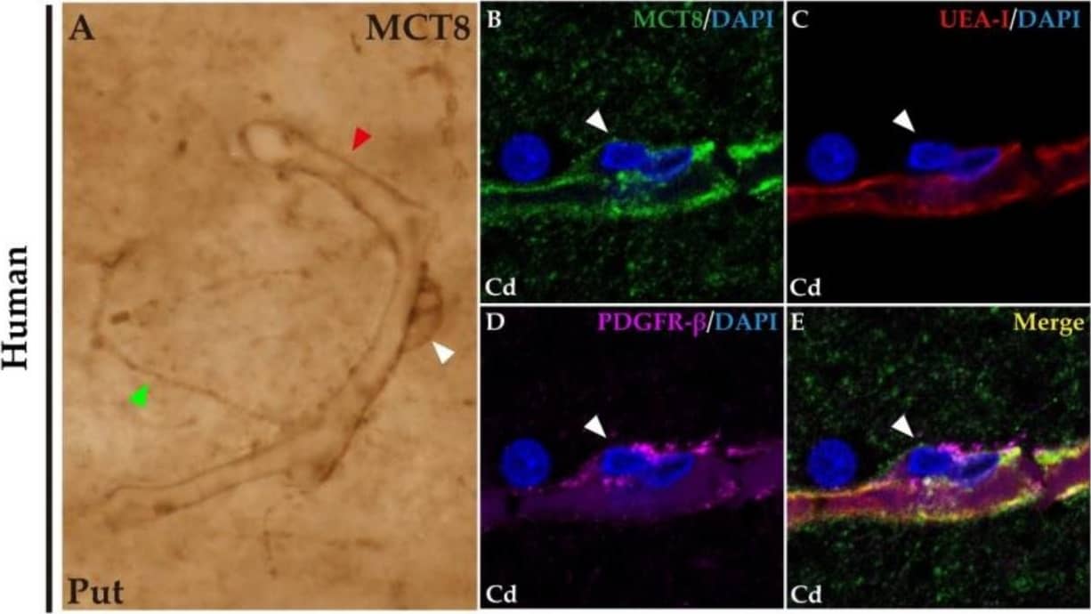
Detection of Human PDGF R beta by Immunohistochemistry Expression of MCT8 and OATP1C1 in blood vessels and brain barriers in the human and macaque basal ganglia and adjacent choroid plexus. (A) Representative brightfield photomicrograph shows immunostaining for MCT8 in the human putamen. Note that an MCT8 immunopositive signal is observed along the capillary wall (red arrowhead), fibers (green arrowhead), and “bump-on-a-log” morphology pericytelike cells (white arrowhead). (B–H) Representative confocal microscope compositions from multiple-stained sections for MCT8 (green), the endothelial marker UEA-I (red), and the vascular and pericyte biomarker PDGFR-beta (purple) in human and macaque caudate nucleus. Merged image (E,H) shows the colocalization of all signals. (B–E) Coexpression of MCT8, UEA-I, and PDGFR-beta is observed in a vessel, while coexpression of MCT8 and PDGFR-beta but not UEA-I is observed in a capillary-associated pericyte (white arrowheads) in humans. (F–H) Coexpression of MCT8 and PDGFR-beta in a vessel and pericytelike cells (white arrowheads) in macaques. Counterstaining with DAPI (blue) shows nuclei of all cells. (I,J) Representative brightfield photomicrographs show immunostaining for MCT8 (I) and OATP1C1 (J) in the macaque choroid plexus at the lateral ventricle. Black arrowheads point to ependymocytes. Cd: caudate nucleus, Put: putamen, PDGFR-beta : platelet-derived growth factor receptor-beta, UEA-I: Ulex europaeus agglutinin-I. Scale bar = 10 μm (A–H) and 50 μm (I,J). Image collected and cropped by CiteAb from the following open publication (https://pubmed.ncbi.nlm.nih.gov/37298594), licensed under a CC-BY license. Not internally tested by R&D Systems.
 View Larger
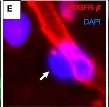
Detection of Mouse PDGF R beta by Immunohistochemistry Pericyte soma on WM capillaries demonstrated by immunofluorescence. A–F. PDGFR‐ beta (red) and COL4 (green) immunofluorescence staining with nuclei (DAPI) (blue). A,B. Low‐ and high‐power images showing capillary segments (arrowheads) with overlapping PDGFR‐ beta (red), COL4 (green) and DAPI (blue). C. Same vessel segment as B with PDGFR‐ beta (red) and COL4 (green); D. COL4 (green) and DAPI (blue); E. PDGFR‐ beta (red) and DAPI (blue). F. Another capillary segment with PDGFR‐ beta (red), COL4 (green) and DAPI (blue) clearly showing pericyte cell body. G–J. Images taken by a confocal microscope showing pericyte cell bodies (arrows) with nuclear stain (DAPI). Capillaries and pericyte processes are revealed by COL4 and PDGFR‐ beta (red) immunoreactivities. Magnification bars: A = 50 µm, F, J = 10 µm. Image collected and cropped by CiteAb from the following open publication (https://pubmed.ncbi.nlm.nih.gov/32705757), licensed under a CC-BY license. Not internally tested by R&D Systems.
Preparation and Storage
- 12 months from date of receipt, -20 to -70 degreesC as supplied. 1 month, 2 to 8 degreesC under sterile conditions after reconstitution. 6 months, -20 to -70 degreesC under sterile conditions after reconstitution.
Background: PDGF R beta
PDGF is a major serum mitogen that can exist as a homo or hetero-dimeric protein consisting of disulfide-linked PDGF-A and PDGF-B chains. The PDGF-AA, PDGF-BB, and PDGF-AB isoforms have been shown to bind to two distinct cell surface PDGF receptors with different affinities. Where as PDGF R alpha binds all three PDGF isoforms with high affinity, PDGF R beta binds PDGF-BB only with high-affinity. Both PDGF R alpha and PDGF R beta are members of the class III subfamily of receptor tyrosine kinases (RTK) that also includes the receptors for M-CSF, SCF, and Flt-3 ligand. All class III RTKs are characterized by the presence of five immunoglobulin-like domains in their extracellular region and a split kinase domain in their intracellular region. PDGF binding induces receptor homo-and hetero-dimerization and signal transduction. The expression of the alpha and beta receptors is independently regulated in various cell types. Recombinant soluble PDGF R beta binds PDGF with high affinity and is potent PDGF antagonist.
- Heldin, C.H. and L. Claesson-Welsh (1994) in Guidebook to Cytokines and Their Receptors, Nicola, N.A. ed. Oxford University Press, New York, p. 202.
Product Datasheets
Citations for Human PDGF R beta Antibody
R&D Systems personnel manually curate a database that contains references using R&D Systems products. The data collected includes not only links to publications in PubMed, but also provides information about sample types, species, and experimental conditions.
62
Citations: Showing 1 - 10
Filter your results:
Filter by:
-
Generation of Human PSC-Derived Kidney Organoids with Patterned Nephron Segments and a De Novo Vascular Network
Authors: JH Low, P Li, EGY Chew, B Zhou, K Suzuki, T Zhang, MM Lian, M Liu, E Aizawa, C Rodriguez, KSM Yong, Q Chen, JM Campistol, M Fang, CC Khor, JN Foo, JC Izpisua Be, Y Xia
Cell Stem Cell, 2019-07-11;0(0):.
-
Intracerebral Hemorrhage-Associated Iron Release Leads to Pericyte-Dependent Cerebral Capillary Function Disruption
Authors: Balk, S;Panier, F;Brandner, S;Coras, R;Blümcke, I;Ekici, AB;Sembill, JA;Schwab, S;Huttner, HB;Sprügel, MI;
Biomolecules
Species: Human
Sample Types: Whole Tissue
Applications: Immunohistochemistry -
Integrating collecting systems in kidney organoids through fusion of distal nephron to ureteric bud
Authors: Shi, M;Crouse, B;Sundaram, N;Shakked, NP;Ester, L;Zhang, W;Janakiram, V;Kopan, R;Helmrath, MA;Bonventre, JV;McCracken, KW;
bioRxiv : the preprint server for biology
Species: Human
Sample Types: Organoid
Applications: Immunocytochemistry -
Kidney organoid models reveal cilium-autophagy metabolic axis as a therapeutic target for PKD both in vitro and in vivo
Authors: Liu, M;Zhang, C;Gong, X;Zhang, T;Lian, MM;Chew, EGY;Cardilla, A;Suzuki, K;Wang, H;Yuan, Y;Li, Y;Naik, MY;Wang, Y;Zhou, B;Soon, WZ;Aizawa, E;Li, P;Low, JH;Tandiono, M;Montagud, E;Moya-Rull, D;Rodriguez Esteban, C;Luque, Y;Fang, M;Khor, CC;Montserrat, N;Campistol, JM;Izpisua Belmonte, JC;Foo, JN;Xia, Y;
Cell stem cell
Species: Human
Sample Types: Organoid
Applications: Immunohistochemistry -
Identification of CCZ1 as an essential lysosomal trafficking regulator in Marburg and Ebola virus infections
Authors: Monteil, V;Kwon, H;John, L;Salata, C;Jonsson, G;Vorrink, SU;Appelberg, S;Youhanna, S;Dyczynski, M;Leopoldi, A;Leeb, N;Volz, J;Hagelkruys, A;Kellner, MJ;Devignot, S;Michlits, G;Foong-Sobis, M;Weber, F;Lauschke, VM;Horn, M;Feldmann, H;Elling, U;Penninger, JM;Mirazimi, A;
Nature communications
Species: Human
Sample Types: Organoids
Applications: IHC -
Mass Spectrometry Reveals that Oxysterols are Secreted from Non-Alcoholic Fatty Liver Disease Induced Organoids
Authors: Kømurcu, KS;Wilhelmsen, I;Thorne, JL;Krauss, S;Wilson, SR;Aizenshtadt, A;Røberg-Larsen, H;
The Journal of steroid biochemistry and molecular biology
Species: Human
Sample Types: Organoid
Applications: Immunohistochemistry -
Thyroid Hormone Transporters MCT8 and OATP1C1 Are Expressed in Projection Neurons and Interneurons of Basal Ganglia and Motor Thalamus in the Adult Human and Macaque Brains
Authors: Wang, T;Wang, Y;Montero-Pedrazuela, A;Prensa, L;Guadaño-Ferraz, A;Rausell, E;
International journal of molecular sciences
Species: Primate - Macaca fascicularis (Crab-eating Monkey or Cynomolgus Macaque), Primate - Macaca mulatta (Rhesus Macaque), Primate - Saimiri sciureus (South American Squirrel Monkey), Human
Sample Types: Whole Tissue
Applications: IHC -
Combinatorial Microgels for 3D ECM Screening and Heterogeneous Microenvironmental Culture of Primary Human Hepatic Stellate Cells
Authors: Ryoo, H;Underhill, GH;
bioRxiv : the preprint server for biology
Species: Human
Sample Types: Whole Cells
Applications: ICC -
Protocol to generate cardiac pericytes from human induced pluripotent stem cells
Authors: Shen M, Zhao SR, Khokhar Y et al.
STAR protocols
-
SARS-CoV-2 triggers pericyte-mediated cerebral capillary constriction
Authors: Chanawee Hirunpattarasilp, Greg James, Jaturon Kwanthongdee, Felipe Freitas, Jiandong Huo, Huma Sethi et al.
Brain
-
NOTCH3 drives meningioma tumorigenesis and resistance to radiotherapy
Authors: Choudhury, A;Cady, M;Lucas, C;Najem, H;Phillips, J;Palikuqi, B;Zakimi, N;Joseph, T;Birrueta, J;Chen, W;Bush, N;Hervey-Jumper, S;Klein, O;Toedebusch, C;Horbinski, C;Magill, S;Bhaduri, A;Perry, A;Dickinson, P;Heimberger, A;Ashworth, A;Crouch, E;Raleigh, D;
bioRxiv
Species: Human
Sample Types: Whole Tissue
Applications: IHC -
Human pericytes degrade diverse alpha-synuclein aggregates
Authors: BV Dieriks, B Highet, A Alik, T Bellande, TJ Stevenson, V Low, TI Park, J Correia, P Schweder, RLM Faull, R Melki, MA Curtis, M Dragunow
PLoS ONE, 2022-11-18;17(11):e0277658.
Species: Human
Sample Types: Whole Cells
Applications: ICC -
Single-cell image analysis reveals over-expression of organic anion transporting polypeptides (OATPs) in human glioblastoma tissue
Authors: Elizabeth Cooper, Zoe Woolf, Molly E V Swanson, Jason Correia, Patrick Schweder, Edward Mee et al.
Neuro-Oncology Advances
-
Evidence of beta amyloid independent small vessel disease in familial Alzheimer's disease
Authors: Jessica Lisa Littau, Lina Velilla, Yoshiki Hase, Nelson David Villalba‐Moreno, Christian Hagel, Dagmar Drexler et al.
Brain Pathology
Species: Human
Sample Types: Whole Tissue
Applications: Immunohistochemistry -
Microglial debris is cleared by astrocytes via C4b-facilitated phagocytosis and degraded via RUBICON-dependent noncanonical autophagy in mice
Authors: T Zhou, Y Li, X Li, F Zeng, Y Rao, Y He, Y Wang, M Liu, D Li, Z Xu, X Zhou, S Du, F Niu, J Peng, X Mei, SJ Ji, Y Shu, W Lu, F Guo, T Wu, TF Yuan, Y Mao, B Peng
Nature Communications, 2022-10-24;13(1):6233.
Species: Mouse
Sample Types: Whole Tissue
Applications: IHC -
miR-181a/b downregulation: a mutation-independent therapeutic approach for inherited retinal diseases
Authors: S Carrella, M Di Guida, S Brillante, D Piccolo, L Ciampi, I Guadagnino, J Garcia Piq, M Pizzo, E Marrocco, M Molinari, G Petrogiann, S Barbato, Y Ezhova, A Auricchio, B Franco, E De Leonibu, EM Surace, A Indrieri, S Banfi
Embo Molecular Medicine, 2022-10-04;0(0):e15941.
Species: Mouse
Sample Types: Whole Tissue
Applications: IHC -
Modeling injury and repair in kidney organoids reveals that homologous recombination governs tubular intrinsic repair
Authors: Gupta N, Matsumoto T, Hiratsuka K et al.
Science Translational Medicine
-
Deubiquitinating enzymes USP4 and USP17 finetune the trafficking of PDGFR beta and affect PDGF-BB-induced STAT3 signalling
Authors: Niki Sarri, Kehuan Wang, Maria Tsioumpekou, Casimiro Castillejo-López, Johan Lennartsson, Carl-Henrik Heldin et al.
Cellular and Molecular Life Sciences
-
PDGF-D−PDGFR beta signaling enhances IL-15–mediated human natural killer cell survival
Authors: Shoubao Ma, Tingting Tang, Xiaojin Wu, Anthony G. Mansour, Ting Lu, Jianying Zhang et al.
Proceedings of the National Academy of Sciences
-
Engagement of the CXCL12-CXCR4 Axis in the Interaction of Endothelial Progenitor Cell and Smooth Muscle Cell to Promote Phenotype Control and Guard Vascular Homeostasis
Authors: SF Mause, E Ritzel, A Deck, F Vogt, EA Liehn
International Journal of Molecular Sciences, 2022-01-14;23(2):.
Species: Human
Sample Types: Whole Cells
Applications: Neutralization -
Pericytes as mediators of infiltration of macrophages in multiple sclerosis
Authors: DK Kaushik, A Bhattachar, BM Lozinski, V Wee Yong
Journal of Neuroinflammation, 2021-12-24;18(1):301.
Species: Human
Sample Types: Whole Tissue
Applications: IHC -
Loss with ageing but preservation of frontal�cortical capillary pericytes in post-stroke dementia, vascular dementia and Alzheimer's disease
Authors: R Ding, Y Hase, M Burke, V Foster, W Stevenson, T Polvikoski, RN Kalaria
Acta neuropathologica communications, 2021-08-02;9(1):130.
Species: Human
Sample Types: Whole Tissue
Applications: IHC -
Retinal capillary degeneration and blood-retinal barrier disruption in murine models of Alzheimer's disease
Authors: H Shi, Y Koronyo, DT Fuchs, J Sheyn, K Wawrowsky, S Lahiri, KL Black, M Koronyo-Ha
Acta Neuropathol Commun, 2020-11-23;8(1):202.
Species: Mouse
Sample Types: Whole Tissue
Applications: IHC -
ShcD Binds DOCK4, Promotes Ameboid Motility and Metastasis Dissemination, Predicting Poor Prognosis in Melanoma
Authors: E Aladowicz, L Granieri, F Marocchi, S Punzi, G Giardina, PF Ferrucci, G Mazzarol, M Capra, G Viale, S Confalonie, S Gandini, F Lotti, L Lanfrancon
Cancers (Basel), 2020-11-13;12(11):.
Species: Human
Sample Types: Cell Lysates
Applications: Western Blot -
Loss of capillary pericytes and the blood–brain barrier in white matter in poststroke and vascular dementias and Alzheimer’s disease
Authors: Ren Ding, Yoshiki Hase, Kamar E. Ameen‐Ali, Michael Ndung'u, William Stevenson, Joseph Barsby et al.
Brain Pathology
Species: Human
Sample Types: Whole Tissue
Applications: Immunohistochemistry -
Reciprocal Interaction between Vascular Filopodia and Neural Stem Cells Shapes Neurogenesis in the Ventral Telencephalon
Authors: B Di Marco, EE Crouch, B Shah, C Duman, MF Paredes, C Ruiz de Al, EJ Huang, J Alfonso
Cell Rep, 2020-10-13;33(2):108256.
Species: Human
Sample Types: Whole Tissue
Applications: IHC -
Cell populations and muscle fiber morphology associated with acute and chronic muscle degeneration in lumbar spine pathology
Authors: Bahar Shahidi, Michael C. Gibbons, Mary Esparza, Vinko Zlomislic, Richard Todd Allen, Steven R. Garfin et al.
JOR SPINE
-
A novel sensitive assay for detection of a biomarker of pericyte injury in cerebrospinal fluid
Authors: Melanie D. Sweeney, Abhay P. Sagare, Maricarmen Pachicano, Michael G. Harrington, Elizabeth Joe, Helena C. Chui et al.
Alzheimer's & Dementia
-
Neutrophil extracellular traps released by neutrophils impair revascularization and vascular remodeling after stroke
Authors: L Kang, H Yu, X Yang, Y Zhu, X Bai, R Wang, Y Cao, H Xu, H Luo, L Lu, MJ Shi, Y Tian, W Fan, BQ Zhao
Nat Commun, 2020-05-19;11(1):2488.
Species: Mouse
Sample Types: Whole Tissue
Applications: IHC -
Identification of early pericyte loss and vascular amyloidosis in Alzheimer’s disease retina
Authors: Haoshen Shi, Yosef Koronyo, Altan Rentsendorj, Giovanna C. Regis, Julia Sheyn, Dieu-Trang Fuchs et al.
Acta Neuropathologica
-
A Practical Guide to the Automated Analysis of Vascular Growth, Maturation and Injury in the Brain
Authors: R Rust, T Kirabali, L Grönnert, B Dogancay, YDP Limasale, A Meinhardt, C Werner, B Laviña, L Kulic, RM Nitsch, C Tackenberg, ME Schwab
Front Neurosci, 2020-03-20;14(0):244.
Species: Human
Sample Types: Whole Tissue
Applications: IHC -
The amyloid-beta degradation intermediate Abeta34 is pericyte-associated and reduced in brain capillaries of patients with Alzheimer's disease
Authors: T Kirabali, S Rigotti, A Siccoli, F Liebsch, A Shobo, C Hock, RM Nitsch, G Multhaup, L Kulic
Acta Neuropathol Commun, 2019-12-03;7(1):194.
Species: Human
Sample Types: Cell Lysates, Whole Cells, Whole Tissue
Applications: ICC, IHC, Western Blot -
Blood brain barrier leakage is not a consistent feature of white matter lesions in CADASIL
Authors: RM Rajani, J Ratelade, V Domenga-De, Y Hase, H Kalimo, RN Kalaria, A Joutel
Acta Neuropathol Commun, 2019-11-21;7(1):187.
Species: Human
Sample Types: Whole Tissue
Applications: IHC-P -
Blood–brain barrier breakdown is an early biomarker of human cognitive dysfunction
Authors: Daniel A. Nation, Melanie D. Sweeney, Axel Montagne, Abhay P. Sagare, Lina M. D’Orazio, Maricarmen Pachicano et al.
Nature Medicine
-
Human blood vessel organoids as a model of diabetic vasculopathy
Authors: Reiner A. Wimmer, Alexandra Leopoldi, Martin Aichinger, Nikolaus Wick, Brigitte Hantusch, Maria Novatchkova et al.
Nature
-
Reductions in brain pericytes are associated with arteriovenous malformation vascular instability
Authors: EA Winkler, H Birk, JK Burkhardt, X Chen, JK Yue, D Guo, WC Rutledge, GF Lasker, C Partow, T Tihan, EF Chang, H Su, H Kim, BP Walcott, MT Lawton
J. Neurosurg., 2018-12-01;0(0):1-11.
Species: Human
Sample Types: Whole Tissue
Applications: IHC-P -
Blood-brain barrier-associated pericytes internalize and clear aggregated amyloid-?42 by LRP1-dependent apolipoprotein E isoform-specific mechanism
Authors: Q Ma, Z Zhao, AP Sagare, Y Wu, M Wang, NC Owens, PB Verghese, J Herz, DM Holtzman, BV Zlokovic
Mol Neurodegener, 2018-10-19;13(1):57.
Species: Human
Sample Types: Whole Tissue
Applications: IHC-P -
Coculture of endothelial progenitor cells and mesenchymal stem cells enhanced their proliferation and angiogenesis through PDGF and Notch signaling
Authors: T Liang, L Zhu, W Gao, M Gong, J Ren, H Yao, K Wang, D Shi
FEBS Open Bio, 2017-10-16;7(11):1722-1736.
Species: Human
Sample Types: Whole Cells
Applications: Neutralization -
Transcriptome analysis of PDGFR?+ cells identifies T-type Ca2+ channel CACNA1G as a new pathological marker for PDGFR?+ cell hyperplasia
Authors: SE Ha, MY Lee, M Kurahashi, L Wei, BG Jorgensen, C Park, PJ Park, D Redelman, KC Sasse, LS Becker, KM Sanders, S Ro
PLoS ONE, 2017-08-14;12(8):e0182265.
Species: Human
Sample Types: Whole Tissue
Applications: IHC-Fr -
A tissue-specific role for intraflagellar transport genes during craniofacial development
Authors: EN Schock, JN Struve, CF Chang, TJ Williams, J Snedeker, AC Attia, RW Stottmann, SA Brugmann
PLoS ONE, 2017-03-27;12(3):e0174206.
Species: Mouse
Sample Types: Whole Tissue
Applications: IHC -
Endothelial Cells from Capillary Malformations Are Enriched for Somatic GNAQ Mutations
Authors: Javier A. Couto, Lan Huang, Matthew P. Vivero, Nolan Kamitaki, Reid A. Maclellan, John B. Mulliken et al.
Plastic and Reconstructive Surgery
-
Shedding of soluble platelet-derived growth factor receptor-beta from human brain pericytes
Authors: Abhay P. Sagare, Melanie D. Sweeney, Jacob Makshanoff, Berislav V. Zlokovic
Neuroscience Letters
-
Immunolocalization of platelet-derived growth factor receptor-beta (PDGFR-beta ) and pericytes in cerebral autosomal dominant arteriopathy with subcortical infarcts and leukoencephalopathy (CADASIL)
Authors: Lucinda J.L. Craggs, Richard Fenwick, Arthur E. Oakley, Masafumi Ihara, Raj N. Kalaria
Neuropathology and Applied Neurobiology
-
Designing and troubleshooting immunopanning protocols for purifying neural cells
Authors: Ben A Barres
Cold Spring Harb Protoc
-
Platelet-derived growth factor primes cancer-associated fibroblasts for apoptosis.
Authors: Rizvi S, Mertens J, Bronk S, Hirsova P, Dai H, Roberts L, Kaufmann S, Gores G
J Biol Chem, 2014-06-27;289(33):22835-49.
Species: Human
Sample Types: Cell Lysates
Applications: Western Blot -
Growth factor receptor-Src-mediated suppression of GRK6 dysregulates CXCR4 signaling and promotes medulloblastoma migration.
Authors: Yuan, Liangpin, Zhang, Hongying, Liu, Jingbo, Rubin, Joshua B, Cho, Yoon-Jae, Shu, Hui Kuo, Schniederjan, Matthew, MacDonald, Tobey J
Mol Cancer, 2013-03-05;12(0):18.
Species: Human
Sample Types: Whole Cells
Applications: Neutralization -
Intrinsic regulation of hemangioma involution by platelet-derived growth factor
Authors: E E Roach, R Chakrabarti, N I Park, E C Keats, J Yip, N G Chan et al.
Cell Death & Disease
-
Tissue transglutaminase promotes PDGF/PDGFR-mediated signaling and responses in vascular smooth muscle cells
Authors: Evgeny A. Zemskov, Irina Mikhailenko, Elizabeth P. Smith, Alexey M. Belkin
Journal of Cellular Physiology
-
PDGFRbeta expression and function in fibroblasts derived from pluripotent cells is linked to DNA demethylation.
J. Cell. Sci., 2012-02-17;125(0):2276-87.
Species: Human
Sample Types: Whole Cells
Applications: Neutralization -
Endothelial Progenitor Cells Induce a Phenotype Shift in Differentiated Endothelial Cells towards PDGF/PDGFR beta Axis-Mediated Angiogenesis
Authors: Moritz Wyler von Ballmoos, Zijiang Yang, Jan Völzmann, Iris Baumgartner, Christoph Kalka, Stefano Di Santo
PLoS ONE
-
Regulation of platelet-derived growth factor receptor function by integrin-associated cell surface transglutaminase.
Authors: Zemskov EA, Loukinova E, Mikhailenko I, Coleman RA, Strickland DK, Belkin AM
J. Biol. Chem., 2009-04-22;284(24):16693-703.
Species: Human
Sample Types: Cell Lysates, Whole Cells
Applications: ICC, Western Blot -
Inhibition of anastomotic intimal hyperplasia using a chimeric decoy strategy against NFkappaB and E2F in a rabbit model.
Authors: Miyake T, Aoki M, Morishita R
Cardiovasc. Res., 2008-05-31;79(4):706-14.
Species: Rabbit
Sample Types: Tissue Homogenates
Applications: Western Blot -
Pancreatic stellate cells: partners in crime with pancreatic cancer cells.
Authors: Vonlaufen A, Joshi S, Qu C, Phillips PA, Xu Z, Parker NR, Toi CS, Pirola RC, Wilson JS, Goldstein D, Apte MV
Cancer Res., 2008-04-01;68(7):2085-93.
Species: Human
Sample Types: Whole Cells
Applications: ICC, Neutralization -
PDGF-C and -D induced proliferation/migration of human RPE is abolished by inflammatory cytokines.
Authors: Li R, Maminishkis A, Wang FE, Miller SS
Invest. Ophthalmol. Vis. Sci., 2007-12-01;48(12):5722-32.
Species: Human
Sample Types: Whole Cells
Applications: Neutralization -
Vascular endothelial growth factor can signal through platelet-derived growth factor receptors.
Authors: Ball SG, Shuttleworth CA, Kielty CM
J. Cell Biol., 2007-04-30;177(3):489-500.
Species: Human
Sample Types: Whole Cells
Applications: Neutralization -
C-KIT, by interacting with the membrane-bound ligand, recruits endothelial progenitor cells to inflamed endothelium.
Authors: Dentelli P, Rosso A, Balsamo A, Colmenares Benedetto S, Zeoli A, Pegoraro M, Camussi G, Pegoraro L, Brizzi MF
Blood, 2007-02-08;109(10):4264-71.
Species: Human
Sample Types: Whole Cells
Applications: Neutralization -
Expression of growth factors and growth factor receptor in non-healing and healing ischaemic ulceration.
Authors: Murphy MO, Ghosh J, Fulford P, Khwaja N, Halka AT, Carter A, Turner NJ, Walker MG
Eur J Vasc Endovasc Surg, 2006-01-20;31(5):516-22.
Species: Human
Sample Types: Whole Tissue
Applications: IHC -
Heterodimerization of FGF-receptor 1 and PDGF-receptor-alpha: a novel mechanism underlying the inhibitory effect of PDGF-BB on FGF-2 in human cells.
Authors: Faraone D, Aguzzi MS, Ragone G, Russo K, Capogrossi MC, Facchiano A
Blood, 2005-12-01;107(5):1896-902.
Species: Human
Sample Types: Whole Cells
Applications: Neutralization -
Decorin inhibition of PDGF-stimulated vascular smooth muscle cell function: potential mechanism for inhibition of intimal hyperplasia after balloon angioplasty.
Authors: Nili N, Cheema AN, Giordano FJ, Barolet AW, Babaei S, Hickey R, Eskandarian MR, Smeets M, Butany J, Pasterkamp G, Strauss BH
Am. J. Pathol., 2003-09-01;163(3):869-78.
Species: Rabbit
Sample Types: Cell Lysates
Applications: Western Blot -
Glucose-induced phosphatidylinositol 3-kinase and mitogen-activated protein kinase-dependent upregulation of the platelet-derived growth factor-beta receptor potentiates vascular smooth muscle cell chemotaxis.
Authors: Campbell M, Allen WE, Silversides JA, Trimble ER
Diabetes, 2003-02-01;52(2):519-26.
Species: Human
Sample Types: Whole Cells
Applications: ICC -
Generation of nephron progenitor cells and kidney organoids from human pluripotent stem cells
Nat Protoc, 2016-12-22;12(1):195-207.
-
SARS-CoV-2 Cell Entry Factors ACE2 and TMPRSS2 Are Expressed in the Microvasculature and Ducts of Human Pancreas but Are Not Enriched in beta Cells
Authors: Coate KC, Cha J, Shrestha S et al.
Int J Mol Sci
FAQs
No product specific FAQs exist for this product, however you may
View all Antibody FAQsReviews for Human PDGF R beta Antibody
Average Rating: 4 (Based on 1 Review)
Have you used Human PDGF R beta Antibody?
Submit a review and receive an Amazon gift card.
$25/€18/£15/$25CAN/¥75 Yuan/¥2500 Yen for a review with an image
$10/€7/£6/$10 CAD/¥70 Yuan/¥1110 Yen for a review without an image
Filter by:


