Human PD-1 Antibody Summary
Accession # Q15116
Applications
Please Note: Optimal dilutions should be determined by each laboratory for each application. General Protocols are available in the Technical Information section on our website.
Scientific Data
 View Larger
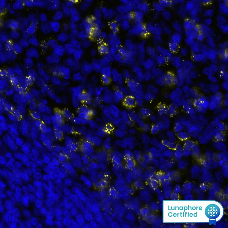
Detection of PD1 in Human Tonsil via seqIF™ staining on COMET™ PD1 Antibody was detected in immersion fixed paraffin-embedded sections of human Tonsil using Rabbit Anti-Human PD1, Monoclonal Antibody (Catalog # MAB11638) at 15ug/mL at 37 ° Celsius for 4 minutes. Before incubation with the primary antibody, tissue underwent an all-in-one dewaxing and antigen retrieval preprocessing using PreTreatment Module (PT Module) and Dewax and HIER Buffer H (pH 9; Epredia Catalog # TA-999-DHBH).Tissue was stained using the Alexa Fluor™ Plus 647 Goat anti-Rabbit IgG Secondary Antibody at 1:200 at 37 ° Celsius for 2 minutes. (Yellow; Lunaphore Catalog # DR647RB) and counterstained with DAPI (blue; Lunaphore Catalog # DR100). Specific staining was localized to the membrane. Protocol available in COMET™ Panel Builder.
 View Larger
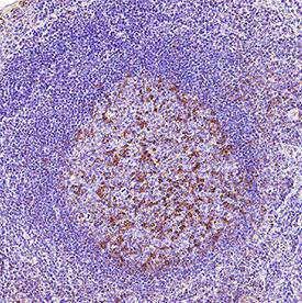
Detection of PD‑1 in Human Tonsil. PD‑1 was detected in immersion fixed paraffin-embedded sections of human tonsil using Rabbit Anti-Human PD‑1 Monoclonal Antibody (Catalog # MAB11638) at 3 µg/ml for 1 hour at room temperature followed by incubation with the Anti-Rabbit IgG VisUCyte™ HRP Polymer Antibody (Catalog # VC003). Before incubation with the primary antibody, tissue was subjected to heat-induced epitope retrieval using VisUCyte Antigen Retrieval Reagent-Basic (Catalog # VCTS021). Tissue was stained using DAB (brown) and counterstained with hematoxylin (blue). Specific staining was localized to the cell surface. View our protocol for IHC Staining with VisUCyte HRP Polymer Detection Reagents.
Preparation and Storage
- 12 months from date of receipt, -20 to -70 °C as supplied.
- 1 month, 2 to 8 °C under sterile conditions after reconstitution.
- 6 months, -20 to -70 °C under sterile conditions after reconstitution.
Background: PD-1
Programmed Death-1 receptor (PD-1), also known as CD279, is type I transmembrane protein belonging to the CD28 family of immune regulatory receptors (1). Other members of this family include CD28, CTLA-4, ICOS, and BTLA (2-5). Mature human PD-1 consists of a 148 amino acid (aa) extracellular region (ECD) with one immunoglobulin-like V-type domain, a 24 aa transmembrane domain, and a 95 aa cytoplasmic region. The human PD-1 ECD shares 65% aa sequence identity with the mouse PD-1 ECD. The cytoplasmic tail contains two tyrosine residues that form the immunoreceptor tyrosine-based inhibitory motif (ITIM) and immunoreceptor tyrosine-based switch motif (ITSM) that are important for mediating PD-1 signaling. PD-1 acts as a monomeric receptor and interacts in a 1:1 stoichiometric ratio with its ligands PD-L1 (B7-H1) and PD-L2 (B7-DC) (6, 7). PD‑1 is expressed on activated T cells, B cells, monocytes, and dendritic cells while PD-L1 expression is constitutive on the same cells and also on nonhematopoietic cells such as lung endothelial cells and hepatocytes (8, 9). Ligation of PD-L1 with PD-1 induces co-inhibitory signals on T cells promoting their apoptosis, anergy, and functional exhaustion (10). Thus, the PD-1: PD-L1 interaction is a key regulator of the threshold of immune response and peripheral immune tolerance (11). Finally, blockade of the PD-1: PD-L1 interaction by either antibodies or genetic manipulation accelerates tumor eradication and shows potential for improving cancer immunotherapy (12, 13, 14).
- Ishida, Y. et al. (1992) EMBO J. 11:3887.
- Sharpe, A.H. and G. J. Freeman (2002) Nat. Rev. Immunol. 2:116.
- Coyle, A. and J. Gutierrez-Ramos (2001) Nat. Immunol. 2:203.
- Nishimura, H. and T. Honjo (2001) Trends Immunol. 22:265.
- Watanabe, N et al. (2003) Nat. Immunol. 4:670.
- Zhang, X. et al. (2004) Immunity 20:337.
- Lázár-Molnár, E. et al. (2008) Proc. Natl. Acad. Sci. USA 105:10483.
- Nishimura, H et al. (1996) Int. Immunol. 8:773.
- Keir, M.E. et al. (2008) Annu. Rev. Immunol. 26:677.
- Butte, M.J. et al. (2007) Immunity 27:111.
- Okazaki, T. et al. (2013) Nat. Immunol. 14:1212.
- Iwai, Y. et al. (2002) Proc. Natl. Acad. Sci. USA 99: 12293.
- Nogrady, B. (2014) Nature 513:S10.
- Swaika, A. et al. (2015) Mol. Immunol. 67:4.
Product Datasheets
FAQs
No product specific FAQs exist for this product, however you may
View all Antibody FAQsReviews for Human PD-1 Antibody
There are currently no reviews for this product. Be the first to review Human PD-1 Antibody and earn rewards!
Have you used Human PD-1 Antibody?
Submit a review and receive an Amazon gift card.
$25/€18/£15/$25CAN/¥75 Yuan/¥2500 Yen for a review with an image
$10/€7/£6/$10 CAD/¥70 Yuan/¥1110 Yen for a review without an image
