Human/Mouse/Rat PKM1/2 Antibody Summary
Ser434-Pro531
Accession # P14618
Applications
Please Note: Optimal dilutions should be determined by each laboratory for each application. General Protocols are available in the Technical Information section on our website.
Scientific Data
 View Larger
Detection of Human, Mouse, and Rat PKM2 by Western Blot. Western blot shows lysates of A549 human lung carcinoma cell line, KG-1 human acute myelogenous leukemia cell line, BaF3 mouse pro-B cell line, and NRK rat normal kidney cell line. PVDF membrane was probed with 0.2 µg/mL of Sheep Anti-Human/Mouse/Rat PKM1/2 Antigen Affinity-purified Polyclonal Antibody (Catalog # AF7244) followed by HRP-conjugated Anti-Sheep IgG Secondary Antibody (HAF016). A specific band was detected for PKM2 at approximately 60 kDa (as indicated). This experiment was conducted under reducing conditions and using Immunoblot Buffer Group 1.
 View Larger
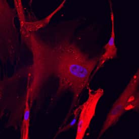
PKM2 in Wi-38 Human Cell Line. PKM2 was detected in immersion fixed Wi-38 human lung fibroblast cell line using Sheep Anti-Human/Mouse/Rat PKM1/2 Antigen Affinity-purified Polyclonal Antibody (Catalog # AF7244) at 10 µg/mL for 3 hours at room temperature. Cells were stained using the NorthernLights™ 557-conjugated Anti-Sheep IgG Secondary Antibody (red; NL010) and counterstained with DAPI (blue). Specific staining was localized to cytoplasm. View our protocol for Fluorescent ICC Staining of Cells on Coverslips.
 View Larger
PKM2 in RAW 264.7 Mouse Cell Line. PKM2 was detected in immersion fixed RAW 264.7 mouse monocyte/macrophage cell line using Sheep Anti-Human/Mouse/Rat PKM1/2 Antigen Affinity-purified Polyclonal Antibody (Catalog # AF7244) at 10 µg/mL for 3 hours at room temperature. Cells were stained using the NorthernLights™ 557-conjugated Anti-Sheep IgG Secondary Antibody (red; NL010) and counterstained with DAPI (blue). Specific staining was localized to cytoplasm. View our protocol for Fluorescent ICC Staining of Cells on Coverslips.
 View Larger
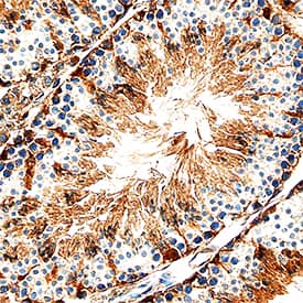
PKM2 in Mouse Testis. PKM2 was detected in perfusion fixed frozen sections of mouse testis using Sheep Anti-Human/Mouse/Rat PKM1/2 Antigen Affinity-purified Polyclonal Antibody (Catalog # AF7244) at 1.7 µg/mL overnight at 4 °C. Tissue was stained using the Anti-Sheep HRP-DAB Cell & Tissue Staining Kit (brown; CTS019) and counterstained with hematoxylin (blue). Specific staining was localized to Sertoli cells in testis. View our protocol for Chromogenic IHC Staining of Frozen Tissue Sections.
 View Larger
PKM2 in Rat Testis. PKM2 was detected in perfusion fixed frozen sections of rat testis using Sheep Anti-Human/Mouse/Rat PKM1/2 Antigen Affinity-purified Polyclonal Antibody (Catalog # AF7244) at 1.7 µg/mL overnight at 4 °C. Tissue was stained using the Anti-Sheep HRP-DAB Cell & Tissue Staining Kit (brown; Catalog # CTS019) and counterstained with hematoxylin (blue). Specific staining was localized to Sertoli cells in testis. View our protocol for Chromogenic IHC Staining of Frozen Tissue Sections.
 View Larger
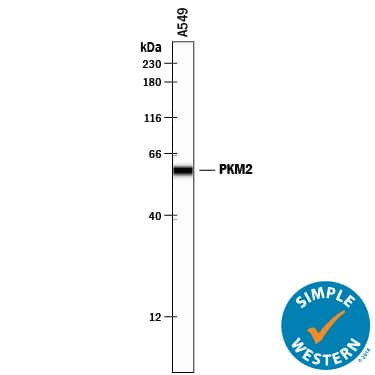
Detection of Human PKM2 by Simple WesternTM. Simple Western lane view shows lysates of A549 human lung carcinoma cell line, loaded at 0.2 mg/mL. A specific band was detected for PKM2 at approximately 60 kDa (as indicated) using 2 µg/mL of Sheep Anti-Human/Mouse/Rat PKM1/2 Antigen Affinity-purified Polyclonal Antibody (Catalog # AF7244) followed by 1:50 dilution of HRP-conjugated Anti-Sheep IgG Secondary Antibody (HAF016). This experiment was conducted under reducing conditions and using the 12-230 kDa separation system.
 View Larger
Western Blot Shows Human PKM2 Specificity by Using Knockout Cell Line. Western blot shows lysates of HeLa human cervical epithelial carcinoma parental cell line and PKM2 knockout HeLa cell line (KO). PVDF membrane was probed with 1 µg/mL of Sheep Anti-Human/Mouse/Rat PKM1/2 Antigen Affinity-purified Polyclonal Antibody (Catalog # AF7244) followed by HRP-conjugated Anti-Sheep IgG Secondary Antibody (HAF016). A specific band was detected for PKM2 at approximately 60 kDa (as indicated) in the parental HeLa cell line, but is not detectable in knockout HeLa cell line. GAPDH (AF5718) is shown as a loading control. This experiment was conducted under reducing conditions and using Immunoblot Buffer Group 1.
 View Larger
Detection of Human PKM2 by Western Blot. Western blot shows lysates of U‑87 MG human glioblastoma/astrocytoma cell line, A549 human lung carcinoma cell line, HeLa human cervical epithelial carcinoma cell line, human skeletal muscle. PVDF membrane was probed with 0.25 µg/mL of Sheep Anti-Human/Mouse/Rat PKM2 Antigen Affinity-purified Polyclonal Antibody (Catalog # AF7244) followed by HRP-conjugated Anti-Sheep IgG Secondary Antibody (HAF016). A specific band was detected for PKM2 at approximately 60 kDa (as indicated). This experiment was conducted under reducing conditions and using Western Blot Buffer Group 1.
Preparation and Storage
- 12 months from date of receipt, -20 to -70 °C as supplied.
- 1 month, 2 to 8 °C under sterile conditions after reconstitution.
- 6 months, -20 to -70 °C under sterile conditions after reconstitution.
Background: PKM2
PKM2 (Pyruvate Kinase isoenzyme M2; also p58, OIP3, THBP1 and CTHBP) is a 58-60 kDa member of the PK family of enzymes. It is widely expressed, being found both intracellularly and in blood, and represents the more common splice variant of the PKM gene. PKM2 generates ATP and pyruvate by catalyzing the transfer of a phosphoryl group from PEP to ADP. Thus, when active, PKM2 promotes energy production and glycolysis. PKM2 exists as a marginally active monomer, with full activity achieved through homotetramerization. Notably, in tumor cells, select oncogenes appear to induce PKM2 homodimerization which limits PKM2 activity. PKM2 is known to be regulated by the binding of T3 and Fru-1,6-bisP. Human PKM2 is 531 amino acids (aa) in length. It contains a catalytic region (aa 43‑527) plus four utilized Ser/Thr and Tyr phosphorylation sites, respectively. PKM1 is another PKM gene splice variant that shows a 45 aa substitution for
aa 389‑433 of PKM2. This variant shows limited expression (striated muscle) and hyperbolic Michaelis-Menten kinetics. There are additional isoform variants of PKM2 that show either a deletion of aa 59‑132, or a 67 aa substitution for aa 1‑82. Over aa 434‑531, human PKM2 shares 95% aa sequence identity with mouse PKM2.
Product Datasheets
Citations for Human/Mouse/Rat PKM1/2 Antibody
R&D Systems personnel manually curate a database that contains references using R&D Systems products. The data collected includes not only links to publications in PubMed, but also provides information about sample types, species, and experimental conditions.
3
Citations: Showing 1 - 3
Filter your results:
Filter by:
-
Integrin B3-PKM2 pathway-mediated aerobic glycolysis contributes to mechanical ventilation-induced pulmonary fibrosis
Authors: Mei S, Xu Q, Hu Y et al.
Theranostics
-
PTEN Protein Phosphatase Activity Is Not Required for Tumour Suppression in the Mouse Prostate
Authors: HM Wise, A Harris, N Kriplani, A Schofield, H Caldwell, MJ Arends, IM Overton, NR Leslie
Biomolecules, 2022-10-19;12(10):.
Species: Mouse
Sample Types: Whole Tissue
Applications: IHC -
The airway epithelium undergoes metabolic reprogramming in individuals at high risk for lung cancer
JCI Insight, 2016-11-17;1(19):e88814.
Species: Human
Sample Types: Cell Lysates
Applications: Western Blot
FAQs
No product specific FAQs exist for this product, however you may
View all Antibody FAQsReviews for Human/Mouse/Rat PKM1/2 Antibody
There are currently no reviews for this product. Be the first to review Human/Mouse/Rat PKM1/2 Antibody and earn rewards!
Have you used Human/Mouse/Rat PKM1/2 Antibody?
Submit a review and receive an Amazon gift card.
$25/€18/£15/$25CAN/¥75 Yuan/¥2500 Yen for a review with an image
$10/€7/£6/$10 CAD/¥70 Yuan/¥1110 Yen for a review without an image

