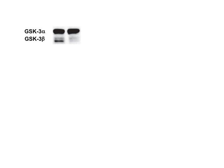
Human/Mouse/Rat GSK-3 alpha / beta Antibody Summary
Accession # P49841
Applications
Please Note: Optimal dilutions should be determined by each laboratory for each application. General Protocols are available in the Technical Information section on our website.
Scientific Data
 View Larger
Detection of Human GSK‑3 alpha / beta by Western Blot. Western blot shows lysates of HeLa human cervical epithelial carcinoma cell line, HT-29 human colon adenocarcinoma cell line, MDA-MB-468 and MCF-7 human breast cancer cell line. PVDF membrane was probed with 0.1 µg/mL of Rabbit Anti-Human/Mouse/ Rat GSK-3a/ beta Antigen Affinity-purified Polyclonal Antibody (Catalog # AF2157) followed by HRP-conjugated Anti-Rabbit IgG Secondary Antibody (Catalog # HAF008). Specific bands were detected for GSK-3a/ beta at approximately 46 and 51 kDa (as indicated). This experiment was conducted under reducing conditions and using Immunoblot Buffer Group 1.
 View Larger
GSK‑3 alpha / beta in HeLa Human Cell Line. GSK-3a/ beta was detected in immersion fixed HeLa human cervical epithelial carcinoma cell line using Rabbit Anti-Human/Mouse/Rat GSK-3a/ beta Antigen Affinity-purified Polyclonal Antibody (Catalog # AF2157) at 10 µg/mL for 3 hours at room temperature. Cells were stained using the NorthernLights™ 557-conjugated Anti-Rabbit IgG Secondary Antibody (red; Catalog # NL004) and counterstained with DAPI (blue). Specific staining was localized to cytoplasm. View our protocol for Fluorescent ICC Staining of Cells on Coverslips.
 View Larger
Detection of GSK‑3 alpha / beta in HeLa Human Cell Line by Flow Cytometry. HeLa human cervical epithelial carcinoma cell line was stained with Rabbit Anti-Human/Mouse/Rat GSK-3a/ beta Antigen Affinity-purified Polyclonal Antibody (Catalog # AF2157, filled histogram) or control antibody (Catalog # AB-105-C, open histogram), followed by Allophycocyanin-conjugated Anti-Rabbit IgG Secondary Antibody (Catalog # F0111). To facilitate intracellular staining, cells were fixed with paraformaldehyde and permeabilized with saponin.
 View Larger
Detection of Mouse GSK-3 alpha/beta by Western Blot GSK3 binds to scaffold formed by histidine-rich cytoplasmic loop domains within the ZIP6-ZIP10 heteromer.(a) Domain organization of ZIP10 and ZIP6 and deletion constructs used in this work to test the hypothesis that a cytoplasmic loop connecting transmembrane (TM) domains III and IV is essential for binding to GSK3 binding. (b) Control Western blot documenting ZIP6 expression products in eluates shown in panel ‘e’. (c) The co-expression of wild-type ZIP6 and ZIP10 proteins comprising intact cytoplasmic loop domains connecting TM domains III and IV is essential for binding of GSK3. GSK3-directed Western blot analysis of cellular lysates and co-immunoprecipitation eluates from in vivo formaldehyde crosslinked cells expressing different combinations of wild-type and/or mutant ZIP6/ZIP10. Note that in addition to protein bands reflecting the expected monomeric molecular weights of GSK3 and ZIP6, the Western blots in panels ‘d’ and ‘e’ revealed the appearance of high molecular weight crosslinked bands, whose appearance depended on the application of the in vivo formaldehyde crosslinking step. Image collected and cropped by CiteAb from the following publication (https://pubmed.ncbi.nlm.nih.gov/28098160), licensed under a CC-BY license. Not internally tested by R&D Systems.
Preparation and Storage
- 12 months from date of receipt, -20 to -70 °C as supplied.
- 1 month, 2 to 8 °C under sterile conditions after reconstitution.
- 6 months, -20 to -70 °C under sterile conditions after reconstitution.
Background: GSK-3 alpha/beta
GSK-3 is a Ser/Thr kinase first identified as an inactivator of Glycogen Synthase. GSK-3 acts as a multifunctional downstream switch that determines the output of numerous signaling pathways. There are two mammalian GSK-3 isoforms encoded by distinct genes, GSK-3 alpha and GSK-3 beta, which are structurally similar, but functionally non-identical. GSK-3a is inhibited by phosphorylation at S21 by Akt and other kinases. GSK-3 alpha and GSK-3 beta share 85% amino acid identity. Dysregulated GSK-3 has been implicated in several diseases including type II diabetes, Alzheimer's disease, bipolar disorder, and cancer.
Product Datasheets
Citations for Human/Mouse/Rat GSK-3 alpha / beta Antibody
R&D Systems personnel manually curate a database that contains references using R&D Systems products. The data collected includes not only links to publications in PubMed, but also provides information about sample types, species, and experimental conditions.
6
Citations: Showing 1 - 6
Filter your results:
Filter by:
-
Single-Cell Spatial MIST for Versatile, Scalable Detection of Protein Markers
Authors: Meah, A;Vedarethinam, V;Bronstein, R;Gujarati, N;Jain, T;Mallipattu, SK;Li, Y;Wang, J;
Biosensors
Species: Mouse
Sample Types: Complex Sample Type
Applications: IHC -
Galectin-3 promotes secretion of proteases that decrease epithelium integrity in human colon cancer cells
Authors: S Li, DM Pritchard, LG Yu
Cell Death & Disease, 2023-04-13;14(4):268.
Species: Human
Sample Types: Cell Lysates
Applications: Western Blot -
A ZIP6-ZIP10 heteromer controls NCAM1 phosphorylation and integration into focal adhesion complexes during epithelial-to-mesenchymal transition
Authors: D Brethour, M Mehrabian, D Williams, X Wang, F Ghodrati, S Ehsani, EA Rubie, JR Woodgett, J Sevalle, Z Xi, E Rogaeva, G Schmitt-Ul
Sci Rep, 2017-01-18;7(0):40313.
Species: Mouse
Sample Types: Cell Lysates
Applications: Immunoprecipitation, Western Blot -
Mycobacteria bypass mucosal NF-kB signalling to induce an epithelial anti-inflammatory IL-22 and IL-10 response.
Authors: Lutay N, Hakansson G, Alaridah N, Hallgren O, Westergren-Thorsson G, Godaly G
PLoS ONE, 2014-01-28;9(1):e86466.
Species: Human
Sample Types: Cell Lysates
Applications: Western Blot -
GSK3beta regulates physiological migration of stem/progenitor cells via cytoskeletal rearrangement.
Authors: Lapid K, Itkin T, D'Uva G, Ovadya Y, Ludin A, Caglio G, Kalinkovich A, Golan K, Porat Z, Zollo M, Lapidot T
J Clin Invest, 2013-03-08;123(4):1705-17.
Species: Mouse
Sample Types: Whole Cells
Applications: Flow Cytometry -
Survival effect of PDGF-CC rescues neurons from apoptosis in both brain and retina by regulating GSK3beta phosphorylation.
Authors: Tang Z, Arjunan P, Lee C, Li Y, Kumar A, Hou X, Wang B, Wardega P, Zhang F, Dong L, Zhang Y, Zhang SZ, Ding H, Fariss RN, Becker KG, Lennartsson J, Nagai N, Cao Y, Li X
J. Exp. Med., 2010-03-15;207(4):867-80.
Species: Rat
Sample Types: Cell Lysates
Applications: Western Blot
FAQs
No product specific FAQs exist for this product, however you may
View all Antibody FAQsReviews for Human/Mouse/Rat GSK-3 alpha / beta Antibody
Average Rating: 4.5 (Based on 4 Reviews)
Have you used Human/Mouse/Rat GSK-3 alpha / beta Antibody?
Submit a review and receive an Amazon gift card.
$25/€18/£15/$25CAN/¥75 Yuan/¥2500 Yen for a review with an image
$10/€7/£6/$10 CAD/¥70 Yuan/¥1110 Yen for a review without an image
Filter by:


