Human/Mouse Cleaved Caspase-3 (Asp175) Antibody Summary
CRGTELDCGIETD
Accession # U26943
Applications
Please Note: Optimal dilutions should be determined by each laboratory for each application. General Protocols are available in the Technical Information section on our website.
Scientific Data
 View Larger
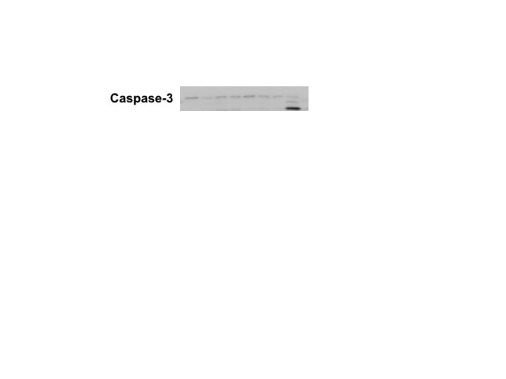
Detection of Human and Mouse Cleaved Caspase‑3 (Asp175) by Western Blot. Western blot shows lysates of Jurkat human acute T cell leukemia cell line and DA3 mouse myeloma cell line untreated (-) or treated (+) with 1 µM staurosporine (STS) for the indicated times. PVDF membrane was probed with 0.5 µg/mL of Human/Mouse Cleaved Caspase-3 (Asp175) Monoclonal Antibody (MAB835), followed by HRP-conjugated Anti-Rabbit IgG Secondary Antibody (Catalog # HAF008). A specific band was detected for Cleaved Caspase-3 (Asp175) at approximately 18 kDa (as indicated). This experiment was conducted under reducing conditions and using Immunoblot Buffer Group 3.
 View Larger
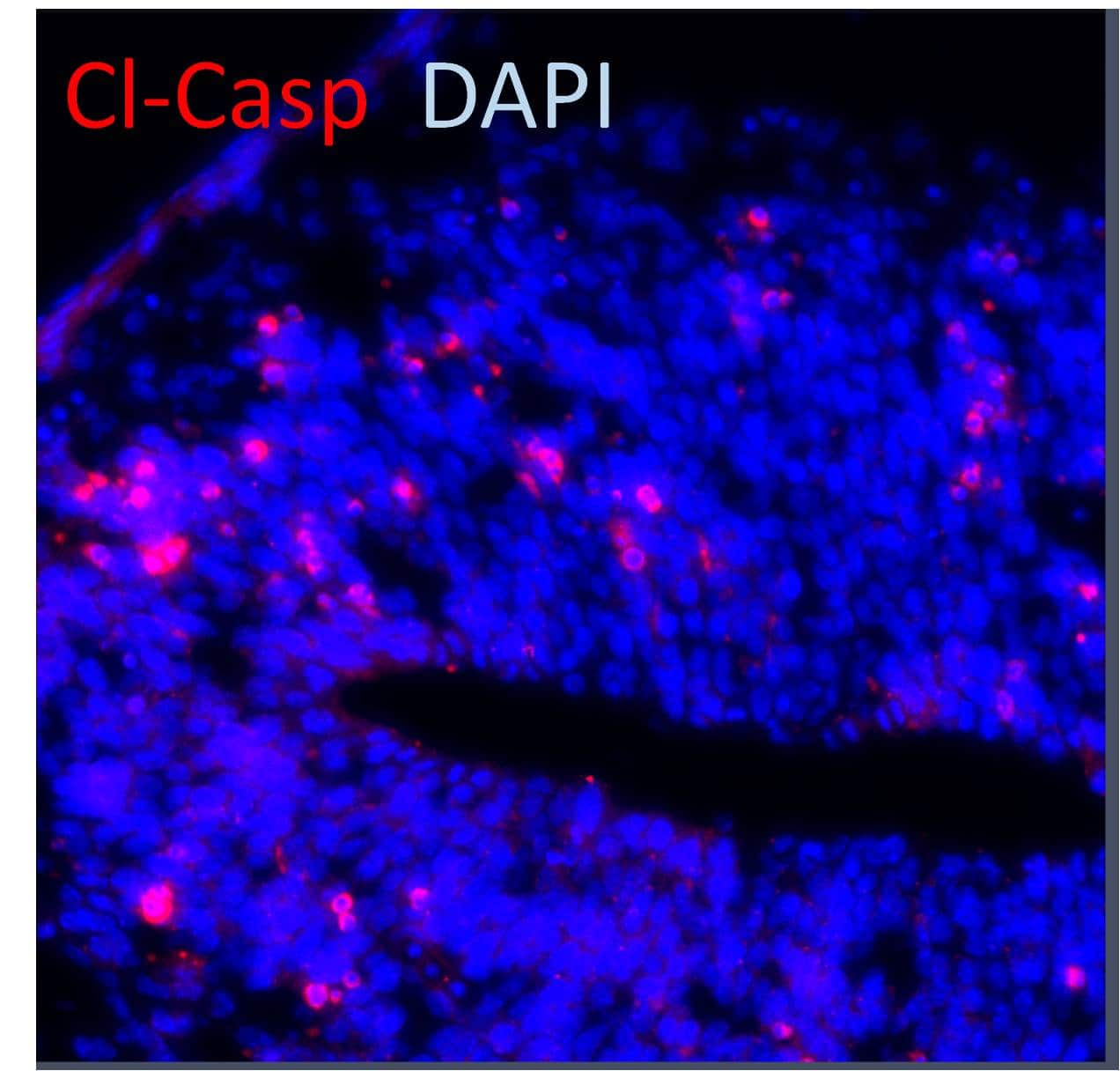
Caspase‑3 in Jurkat Human Cell Line. Caspase-3 was detected in immersion fixed Jurkat human acute T cell leukemia cell line treated with staurosporin using Human/Mouse Cleaved Caspase-3 (Asp175) Monoclonal Antibody (Catalog # MAB835) at 10 µg/mL for 3 hours at room temperature. Cells were stained using the NorthernLights™ 557-conjugated Anti-Rabbit IgG Secondary Antibody (red; Catalog # NL004) and counterstained with DAPI (blue). View our protocol for Fluorescent ICC Staining of Non-adherent Cells.
 View Larger
Caspase‑3 in Human Colon Cancer Tissue. Caspase-3 was detected in immersion fixed paraffin-embedded sections of human colon cancer tissue using Rabbit Anti-Human/Mouse Cleaved Caspase-3 (Asp175) Monoclonal Antibody (Catalog # MAB835) at 0.3 µg/mL for 1 hour at room temperature followed by incubation with the Anti-Rabbit IgG VisUCyte™ HRP Polymer Antibody (Catalog # VC003). Before incubation with the primary antibody, tissue was subjected to heat-induced epitope retrieval using Antigen Retrieval Reagent-Basic (Catalog # CTS013). Tissue was stained using DAB (brown) and counterstained with hematoxylin (blue). Specific staining was localized to cytoplasm. View our protocol for IHC Staining with VisUCyte HRP Polymer Detection Reagents.
 View Larger
Detection of Cleaved Caspase‑3 in Jurkat Human Cell Line by Flow Cytometry. Jurkat human acute T cell leukemia cell line untreated (open histogram) or treated with 3 µM Staurosporine for 3 hours (filled histogram) was stained with Rabbit Anti-Human/Mouse Caspase-3 Monoclonal Antibody (Catalog # MAB835, filled histogram) followed by anti-Rabbit IgG FITC-conjugated secondary antibody (Catalog # F0112). To facilitate intracellular staining, cells were fixed with Flow Cytometry Fixation Buffer (Catalog # FC004) and permeabilized with 90% methanol. View our protocol for Staining Intracellular Molecules.
 View Larger
Detection of Human Caspase-3 by Immunocytochemistry/Immunofluorescence Caspase-3 activation and actin cytoskeletal organization in melanoma A375 cells cultured in the presence of PFII.Cells were grown on glass coverslips in the presence of 65, 130, 260 µM or 1 µM staurosporine (STS). (A–E) Active caspase-3 was visualized with anti-active caspase-3 antibody followed by a FITC-conjugated secondary antibody (green). (F–J) Actin was visualized using laser scanning confocal microscope (LSCM) after staining with Alexa Fluor 568 - conjugated phalloidin (red). Scale bar - 50 µm. Image collected and cropped by CiteAb from the following publication (https://dx.plos.org/10.1371/journal.pone.0057991), licensed under a CC-BY license. Not internally tested by R&D Systems.
 View Larger
Detection of Mouse Caspase-3 by Western Blot Epidermis-specific deletion of Rac1 increases UV-light-induced keratinocyte apoptosis in vivo. (a) H/E staining of UV-irradiated skin of Rac1 fl/fl and Rac1-EKO mice at 12 h after UV-irradiation. Black arrows indicate sunburn cells. (b) Graph shows the percentage number of sunburn cells at 12 h with (red bars) or without (blue bars) UV-irradiation in Rac1 fl/fl (n=4) and Rac1-EKO (n=5) mice. The percentage of sunburn cells within the epidermis after UV-irradiation in Rac1 fl/fl mice was 3.4%, whereas in Rac1-EKO mice it was 8.6% of total epidermal keratinocytes. Non-irradiated samples showed <1% sunburn cells in both the genotypes. (c) Immunostainings against cleaved caspase-3 (green) of UV-irradiated skin of Rac1 fl/fl and Rac1-EKO mice at 12 h after UV-irradiation. Nuclei are stained in blue. Scale bar=100 μm. (d) Western blot analysis of cleaved caspase-3 from epidermal lysates of untreated (no UV) and UV-light treated (UV) Rac1 fl/fl and Rac1-EKO mice. Non-irradiated controls showed no bands for cleaved capsase-3, whereas samples of irradiated epidermis showed cleaved caspase-3-specific bands at ~25 kDa and 23 kDa. Numbers on the left denote molecular weights in kDa. (e) Graph shows densitometry analysis of western blot in (d). Error bars show S.D. Asterisks show P-value<0.001 Image collected and cropped by CiteAb from the following publication (https://pubmed.ncbi.nlm.nih.gov/28277539), licensed under a CC-BY license. Not internally tested by R&D Systems.
 View Larger
Detection of Human Caspase-3 by Immunocytochemistry/Immunofluorescence Apoptotic markers in PFII treated NHDFs.(A–C) [Ca2+]i fluorescence visualizations in cells as revealed by using an LSCM with fluorescent probe Fluo3/AM, scale bar - 100 µm. (D–F) Externalized phosphatidylserine by annexin V-fluorescein binding after PFII treatment, scale bar - 100 µm. Third (G–I) and fourth (J–L) panels show actin cytoskeletal organization and caspase-3 activation. Scale bar - 50 µm. Image collected and cropped by CiteAb from the following publication (https://dx.plos.org/10.1371/journal.pone.0057991), licensed under a CC-BY license. Not internally tested by R&D Systems.
 View Larger
Detection of Mouse Caspase-3 by Western Blot Rac1 deficiency increases sensitivity towards UV-light-induced keratinocyte apoptosis in vitro. (a and b) Western blot analysis of cleaved caspase-3 from Rac1 fl/fl and Rac1-EKO cultured keratinocytes at 6 h (a) and at 12 h (b) with (UV) or without (no UV) UV-irradiation. (c) Densitometry analysis of cleaved caspase-3 at 6 h after UV-irradiation from Rac1 fl/fl and Rac1-EKO keratinocytes. (d) Quantification of CPDs by CPD ELISA carried out from genomic DNA isolated from Rac1 fl/fl and Rac1-EKO mouse primary keratinocytes immediately after UV-irradiation. Error bars show S.D. Asterisks show P-value<0.01 Image collected and cropped by CiteAb from the following publication (https://pubmed.ncbi.nlm.nih.gov/28277539), licensed under a CC-BY license. Not internally tested by R&D Systems.
 View Larger
Detection of Mouse Caspase-3 by Western Blot Increase in UV-light-induced apoptosis in Rac1-deficient keratinocytes requires activation of caspase-8. (a) Western blot analysis of cleaved caspase-8 from Rac1 fl/fl and Rac1-EKO cultured keratinocytes at 6 h with (UV) or without (no UV) UV-irradiation. (b) Densitometry analysis of fold change of cleaved caspase-8 in (a) normalized to GAPDH in Rac1 fl/fl and Rac1-EKO samples after UV-irradiation. (c) Western blot analysis of cleaved caspase-8 from CPDPL/Rac1-EKO epidermal lysates from mice kept in the dark or under the photoreactivtion lamp (PR). (d) Densitometric analysis of cleaved caspase-8 western blots in c. Error bars show S.D. (e) Western blot analysis of cleaved caspase-3 from wild-type (WT) and TNF receptor-1-deficient (TNFR-1 KO) cultured keratinocytes at 6 h with (UV) or without (no UV) UV-irradiation. (f) Western blot analysis of cleaved caspase-3 from TNFR-1 KO cultured keratinocytes incubated with DMSO or Rac1 inhibitor (EHT 1864) at 6 h with (UV) or without (no UV) UV-irradiation. GAPDH is used as a loading control. Numbers on the left denote molecular weights in kDa. * and *** represent P-value<0.05 and <0.001, respectively Image collected and cropped by CiteAb from the following publication (https://pubmed.ncbi.nlm.nih.gov/28277539), licensed under a CC-BY license. Not internally tested by R&D Systems.
 View Larger
Detection of Rat Caspase-3 by Western Blot Western blot analysis of high-mobility group box-1 (HMGB1), brain-derived neurotrophic factor (BDNF), synaptophysin, and cleaved caspase-3 in rat retinas. There is a significant increase in the expression of HMGB1 and cleaved caspase-3 and a significant decrease in the expression of BDNF and synaptophysin in the retinas of diabetic rats (D) compared with the nondiabetic control rats (N). Each experiment was repeated 2 to 3 times with fresh samples (n = 6). Image collected and cropped by CiteAb from the following publication (https://pubmed.ncbi.nlm.nih.gov/23766563), licensed under a CC-BY license. Not internally tested by R&D Systems.
 View Larger
Detection of Human Caspase-3 by Immunocytochemistry/Immunofluorescence BFSE decreases macrophage viability. THP-1 macrophage, or MDM cells, were exposure to air control or 1%, 2.5%, 5%, 10% BFSE or 10% CSE for 24 h. LDH from THP-1 macrophage or MDM cells was measured in supernatants after 24 h (n = 4) (A). THP-1 macrophage expression of Bcl-2 and PARP was assessed by western blot (B), band densitometry analysis of Bcl-2 (C) and PARP (D) was performed, data is a representation of three independent experiments, and was baselined to the air treated control sample, and normalized to beta -actin expression. Error bars represent 95% confidence intervals. Representative confocal images of active caspase-3 (E) and PAR (F), in THP-1 macrophage with quantitative MFI measurement of active caspase-3 (G) and PAR (H). (n = 4) *p < 0.05, **p < 0.01. Image collected and cropped by CiteAb from the following publication (https://pubmed.ncbi.nlm.nih.gov/30194323), licensed under a CC-BY license. Not internally tested by R&D Systems.
 View Larger
Detection of Human Caspase-3 by Western Blot Rac1 deficiency increases sensitivity towards UV-light-induced keratinocyte apoptosis in vitro. (a and b) Western blot analysis of cleaved caspase-3 from Rac1 fl/fl and Rac1-EKO cultured keratinocytes at 6 h (a) and at 12 h (b) with (UV) or without (no UV) UV-irradiation. (c) Densitometry analysis of cleaved caspase-3 at 6 h after UV-irradiation from Rac1 fl/fl and Rac1-EKO keratinocytes. (d) Quantification of CPDs by CPD ELISA carried out from genomic DNA isolated from Rac1 fl/fl and Rac1-EKO mouse primary keratinocytes immediately after UV-irradiation. Error bars show S.D. Asterisks show P-value<0.01 Image collected and cropped by CiteAb from the following publication (https://pubmed.ncbi.nlm.nih.gov/28277539), licensed under a CC-BY license. Not internally tested by R&D Systems.
 View Larger
Detection of Human Caspase-3 by Immunocytochemistry/Immunofluorescence Caspase-3 activation and actin cytoskeletal organization in melanoma A375 cells cultured in the presence of PFII.Cells were grown on glass coverslips in the presence of 65, 130, 260 µM or 1 µM staurosporine (STS). (A–E) Active caspase-3 was visualized with anti-active caspase-3 antibody followed by a FITC-conjugated secondary antibody (green). (F–J) Actin was visualized using laser scanning confocal microscope (LSCM) after staining with Alexa Fluor 568 - conjugated phalloidin (red). Scale bar - 50 µm. Image collected and cropped by CiteAb from the following publication (https://dx.plos.org/10.1371/journal.pone.0057991), licensed under a CC-BY license. Not internally tested by R&D Systems.
 View Larger
Detection of Human Caspase-3 by Immunocytochemistry/Immunofluorescence Caspase-3 activation and actin cytoskeletal organization in melanoma A375 cells cultured in the presence of PFII.Cells were grown on glass coverslips in the presence of 65, 130, 260 µM or 1 µM staurosporine (STS). (A–E) Active caspase-3 was visualized with anti-active caspase-3 antibody followed by a FITC-conjugated secondary antibody (green). (F–J) Actin was visualized using laser scanning confocal microscope (LSCM) after staining with Alexa Fluor 568 - conjugated phalloidin (red). Scale bar - 50 µm. Image collected and cropped by CiteAb from the following publication (https://dx.plos.org/10.1371/journal.pone.0057991), licensed under a CC-BY license. Not internally tested by R&D Systems.
 View Larger
Detection of Rat Caspase-3 by Western Blot Western blot analysis of rat retinas. Intravitreal administration of high-mobility group box-1 (HMGB1) induced a significant upregulation of the expression of HMGB1 and cleaved caspase-3 and a significant downregulation of the expression of brain-derived neurotrophic factor (BDNF) and synaptophysin compared with intravitreal administration of phosphate buffer saline (PBS). Each experiment was repeated 2 to 3 times with fresh samples (n = 6). Image collected and cropped by CiteAb from the following publication (https://pubmed.ncbi.nlm.nih.gov/23766563), licensed under a CC-BY license. Not internally tested by R&D Systems.
 View Larger
Detection of Human Caspase-3 by Immunocytochemistry/Immunofluorescence Apoptotic markers in PFII treated NHDFs.(A–C) [Ca2+]i fluorescence visualizations in cells as revealed by using an LSCM with fluorescent probe Fluo3/AM, scale bar - 100 µm. (D–F) Externalized phosphatidylserine by annexin V-fluorescein binding after PFII treatment, scale bar - 100 µm. Third (G–I) and fourth (J–L) panels show actin cytoskeletal organization and caspase-3 activation. Scale bar - 50 µm. Image collected and cropped by CiteAb from the following publication (https://dx.plos.org/10.1371/journal.pone.0057991), licensed under a CC-BY license. Not internally tested by R&D Systems.
 View Larger
Detection of Human Caspase-3 by Immunocytochemistry/Immunofluorescence Caspase-3 activation and actin cytoskeletal organization in melanoma A375 cells cultured in the presence of PFII.Cells were grown on glass coverslips in the presence of 65, 130, 260 µM or 1 µM staurosporine (STS). (A–E) Active caspase-3 was visualized with anti-active caspase-3 antibody followed by a FITC-conjugated secondary antibody (green). (F–J) Actin was visualized using laser scanning confocal microscope (LSCM) after staining with Alexa Fluor 568 - conjugated phalloidin (red). Scale bar - 50 µm. Image collected and cropped by CiteAb from the following publication (https://dx.plos.org/10.1371/journal.pone.0057991), licensed under a CC-BY license. Not internally tested by R&D Systems.
 View Larger
Detection of Mouse Caspase-3 by Western Blot Increase in UV-light-induced apoptosis in Rac1-deficient keratinocytes requires activation of caspase-8. (a) Western blot analysis of cleaved caspase-8 from Rac1 fl/fl and Rac1-EKO cultured keratinocytes at 6 h with (UV) or without (no UV) UV-irradiation. (b) Densitometry analysis of fold change of cleaved caspase-8 in (a) normalized to GAPDH in Rac1 fl/fl and Rac1-EKO samples after UV-irradiation. (c) Western blot analysis of cleaved caspase-8 from CPDPL/Rac1-EKO epidermal lysates from mice kept in the dark or under the photoreactivtion lamp (PR). (d) Densitometric analysis of cleaved caspase-8 western blots in c. Error bars show S.D. (e) Western blot analysis of cleaved caspase-3 from wild-type (WT) and TNF receptor-1-deficient (TNFR-1 KO) cultured keratinocytes at 6 h with (UV) or without (no UV) UV-irradiation. (f) Western blot analysis of cleaved caspase-3 from TNFR-1 KO cultured keratinocytes incubated with DMSO or Rac1 inhibitor (EHT 1864) at 6 h with (UV) or without (no UV) UV-irradiation. GAPDH is used as a loading control. Numbers on the left denote molecular weights in kDa. * and *** represent P-value<0.05 and <0.001, respectively Image collected and cropped by CiteAb from the following publication (https://pubmed.ncbi.nlm.nih.gov/28277539), licensed under a CC-BY license. Not internally tested by R&D Systems.
 View Larger
Detection of Mouse Human/Mouse Cleaved Caspase-3 (Asp175) Antibody by Western Blot Gamma herpesvirus 68 ( gamma HV68) replicates in alveolar epithelial cells (AECs). (A) Wild-type mice were infected with 5 × 10⁴ PFU gamma HV68 on day 0. On day 7 after infection, frozen sections were prepared, and stained with a rabbit polyclonal antisera against gamma HV68, or with non-immune rabbit sera as control. The goat anti-rabbit secondary was linked to alkaline phosphatase. Vivid replication of gamma HV68 is visible in alveolar lining cells (original magnification × 100). Sections shown are representative of four mice examined. (B) AECs were isolated from lungs of Balb/c or TLR-9-/- mice treated with bleomycin plus gamma HV68 on day 21, and were cultured on fibronectin-coated slides (TiterTek). Sections were stained with antibodies (M30 Cytodeath), and the number of positive cells per high power field (HPF; ×400) were calculated (n = 30 HPF per genotype). (C) AECs were isolated from Balb/c or TLR-9-/- mice and cells were infected in vitro with 0.01 or 0.001 PFU gamma HV68 for 48 hours. Cell lysates were then analyzed for cleaved caspase 3 by western blotting. Data are from one experiment, representative of two. Image collected and cropped by CiteAb from the following publication (https://pubmed.ncbi.nlm.nih.gov/21810214), licensed under a CC-BY license. Not internally tested by R&D Systems.
 View Larger
Detection of Human Human/Mouse Cleaved Caspase-3 (Asp175) Antibody by Immunohistochemistry Assessment of the skin structure and apoptosis upon treatment with wound gels. Representative images and zoom-ins of the boxed areas of haematoxylin and eosin (H&E)-stained tape-stripped (TS) sections from biopsies (age 38 years) either left untreated or treated with indicated wound gels using a light microscope. The presence of caspase-3 expressing cells was evaluated using a caspase-3 (CASP3) antibody and visualisation with a secondary Alexa Fluor™ 546 AB (red). Nuclei were counterstained with DAPI (blue). Scale bar = 50 μm. Image collected and cropped by CiteAb from the following publication (https://pubmed.ncbi.nlm.nih.gov/36261541), licensed under a CC-BY license. Not internally tested by R&D Systems.
Preparation and Storage
- 12 months from date of receipt, -20 to -70 °C as supplied.
- 1 month, 2 to 8 °C under sterile conditions after reconstitution.
- 6 months, -20 to -70 °C under sterile conditions after reconstitution.
Background: Caspase-3
Caspase-3 (Cysteine-aspartic acid protease 3/Casp3; also Yama, apopain and CPP32) is a 29 kDa heterodimer that belongs to the peptidase C14A family of enzymes. It is widely expressed and considered to be the major executioner caspase in the apoptotic cascade. Human procaspase-3 is a 32 kDa, 277 amino acid (aa) protein and is normally an inactive homodimer. Following cell stress/activation, procaspase-3 undergoes proteolysis to generate an N-terminal 148 aa p17/17 kDa subunit (aa 29-175), plus a 102 aa C-terminal p12/12 kDa subunit. These subunits noncovalently heterodimerize, and associate with another p17/p12 heterodimer to form an active enzyme. There is one potential variant that shows an alternative start site nine aa upstream of the standard start site coupled with a 21 aa substitution for aa 162-277. Over aa 29-175, human and mouse caspase-3 share 87% aa identity.
Product Datasheets
Citations for Human/Mouse Cleaved Caspase-3 (Asp175) Antibody
R&D Systems personnel manually curate a database that contains references using R&D Systems products. The data collected includes not only links to publications in PubMed, but also provides information about sample types, species, and experimental conditions.
99
Citations: Showing 1 - 10
Filter your results:
Filter by:
-
ER Redox Homeostasis Regulates Proinsulin Trafficking and Insulin Granule Formation in the Pancreatic Islet beta-Cell
Authors: Rohli KE, Boyer CK, Bearrows SC et al.
Function (Oxford, England)
-
Impaired mitochondrial fatty acid synthesis leads to neurodegeneration in mice.
Authors: Nair RR, koivisto H, Jokivarsi k et al.
J. Neurosci.
-
Downregulation of CDC20 Increases Radiosensitivity through Mcl-1/p-Chk1-Mediated DNA Damage and Apoptosis in Tumor Cells
Authors: Yang Gao, Pengbo Wen, Bin Chen, Guanshuo Hu, Lijun Wu, An Xu et al.
International Journal of Molecular Sciences
-
Unconventional Secretion of Adipocyte Fatty Acid Binding Protein 4 Is Mediated By Autophagic Proteins in a Sirtuin-1–Dependent Manner
Authors: Ajeetha Josephrajan, Ann V. Hertzel, Ellie K. Bohm, Michael W. McBurney, Shin-Ichiro Imai, Douglas G. Mashek et al.
Diabetes
-
DL-3-n-butylphthalide protects endothelial cells against advanced glycation end product-induced injury by attenuating oxidative stress and inflammation responses
Authors: Chang-Yun Liu, Zhen-Hua Zhao, Zhi-Ting Chen, Chun-Hui Che, Zhang-Yu Zou, Xiao-Min Wu et al.
Experimental and Therapeutic Medicine
-
Augmin deficiency in neural stem cells causes p53-dependent apoptosis and aborts brain development
Authors: Ricardo Viais, Marcos Fariña-Mosquera, Marina Villamor-Payà, Sadanori Watanabe, Lluís Palenzuela, Cristina Lacasa et al.
eLife
-
Knockdown of anti-silencing function 1B histone chaperone induces cell apoptosis via repressing PI3K/Akt pathway in prostate cancer
Authors: Guangye Han, Xinjun Zhang, Pei Liu, Quanfeng Yu, Zeyu Li, Qinnan Yu et al.
International Journal of Oncology
-
Knockdown of RRM1 in tumor cells promotes radio-/chemotherapy induced ferroptosis by regulating p53 ubiquitination and p21-GPX4 signaling axis
Authors: Yang Gao, Bin Chen, Ruru Wang, An Xu, Lijun Wu, Huayi Lu et al.
Cell Death Discovery
-
Endoplasmic reticulum stress and IRE-1 signaling cause apoptosis in colon cancer cells in response to andrographolide treatment
Authors: Aditi Banerjee, Hafiz Ahmed, Peixin Yang, Steven J. Czinn, Thomas G. Blanchard
Oncotarget
-
Intercellular Adhesion Molecule-1-Induced Posttraumatic Brain Injury Neuropathology in the Prefrontal Cortex and Hippocampus Leads to Sensorimotor Function Deficits and Psychological Stress
Authors: Bhowmick S, Malat A, Caruso D Et Al.
eNeuro
-
Impact of Andrographolide and Melatonin Combinatorial Drug Therapy on Metastatic Colon Cancer Cells and Organoids
Authors: Neha Sharda, Tamaki Ikuse, Elizabeth Hill, Sonia Garcia, Steven J Czinn, Andrea Bafford et al.
Clin Med Insights Oncol
-
Smac-mimetic enhances antitumor effect of standard chemotherapy in ovarian cancer models via Caspase 8-independent mechanism
Authors: Lidia F. Hernandez, Angie B. Dull, Soumya Korrapati, Christina M. Annunziata
Cell Death Discovery
-
Preclinical evaluation of the first intravenous small molecule MDM2 antagonist alone and in combination with temozolomide in neuroblastoma
Authors: Lindi Chen, Fabio Pastorino, Philip Berry, Jennifer Bonner, Calum Kirk, Katrina M. Wood et al.
International Journal of Cancer
-
Dysfunctional SEMA3E signaling underlies gonadotropin-releasing hormone neuron deficiency in Kallmann syndrome
Authors: Anna Cariboni, Valentina André, Sophie Chauvet, Daniele Cassatella, Kathryn Davidson, Alessia Caramello et al.
Journal of Clinical Investigation
-
High-Mobility Group Box-1 Induces Decreased Brain-Derived Neurotrophic Factor-Mediated Neuroprotection in the Diabetic Retina
Authors: Ahmed M. Abu El-Asrar, Mohd Imtiaz Nawaz, Mohammad Mairaj Siddiquei, Abdullah S. Al-Kharashi, Dustan Kangave, Ghulam Mohammad
Mediators of Inflammation
-
Effect of brefeldin A and castanospermine on resistant cell lines as supplements in anticancer therapy
Authors: KAROLINA WOJTOWICZ, RADOSŁAW JANUCHOWSKI, PATRYCJA SOSIŃSKA, MICHAŁ NOWICKI, MACIEJ ZABEL
Oncology Reports
-
Epidermal Rac1 regulates the DNA damage response and protects from UV-light-induced keratinocyte apoptosis and skin carcinogenesis
Authors: J Deshmukh, R Pofahl, I Haase
Cell Death Dis, 2017-03-09;8(3):e2664.
-
Intratumoral injection of pEGFC1-IGFBP7 inhibits malignant melanoma growth in C57BL/6J mice by inducing apoptosis and down-regulating VEGF expression
Authors: Rong-Yi Chen, Hong-Xiang Chen, Ping Jian, Li Xu, Juan Li, Yi-Ming Fan et al.
Oncology Reports
-
Development of a quantitative pharmacodynamic assay for apoptosis in fixed tumor tissue and its application in distinguishing cytotoxic drug—induced DNA double strand breaks from DNA double strand breaks associated with apoptosis
Authors: Angie B. Dull, Deborah Wilsker, Melinda Hollingshead, Christina Mazcko, Christina M. Annunziata, Amy K. LeBlanc et al.
Oncotarget
-
A piRNA-like Small RNA Induces Chemoresistance to Cisplatin-Based Therapy by Inhibiting Apoptosis in Lung Squamous Cell Carcinoma
Authors: Yuyan Wang, Tyler Gable, Mark Z. Ma, David Clark, Jun Zhao, Yi Zhang et al.
Molecular Therapy - Nucleic Acids
-
Notch1 is required to maintain supporting cell identity and vestibular function during maturation of the mammalian balance organs
Authors: Heffer, A;Lee, C;Holt, JC;Kiernan, AE;
bioRxiv : the preprint server for biology
Species: Mouse
Sample Types: Whole Tissue
Applications: Immunohistochemistry -
Interferon-? Overexpression in Adipose Tissue-Derived Stem Cells Induces HepG2 and Macrophage Cell Death in Liver Tumor Organoids via Induction of TNF-Related Apoptosis-Inducing Ligand Expression
Authors: Yoon, Y;Kim, CW;Kim, MY;Baik, SK;Jung, PY;Eom, YW;
International journal of molecular sciences
Species: Human
Sample Types: Organoids
Applications: Western Blot, IHC -
Improving anti-tumor efficacy of low-dose Vincristine in rhabdomyosarcoma via the combination therapy with FOXM1 inhibitor RCM1
Authors: Donovan J, Deng Z, Bian F et al.
Frontiers in oncology
-
CCR3 blockage elicits polyploidization associated with the signatures of epithelial-mesenchymal transition in carcinoma cell lines
Authors: Y Kaibori, D Nagakubo
Cancer Gene Therapy, 2022-09-19;0(0):.
Species: Human
Sample Types: Whole Cells
Applications: ICC -
Cellular recovery after prolonged warm ischaemia of the whole body
Authors: D Andrijevic, Z Vrselja, T Lysyy, S Zhang, M Skarica, A Spajic, D Dellal, SL Thorn, RB Duckrow, S Ma, PQ Duy, AU Isiktas, D Liang, M Li, SK Kim, SG Daniele, K Banu, S Perincheri, MC Menon, A Huttner, KN Sheth, KT Gobeske, GT Tietjen, HP Zaveri, SR Latham, AJ Sinusas, N Sestan
Nature, 2022-08-03;608(7922):405-412.
Species: Human
Sample Types: Whole Tissue
Applications: IHC -
Insights into the Pharmacokinetics and In Vitro Cell-Based Studies of the Imidazoline I2 Receptor Ligand B06
Authors: A Bagán, JA Morales-Ga, C Griñán-Fer, C Díaz, J Pérez Del, MC Ramos, F Vicente, B Pérez, J Brea, MI Loza, M Pallàs, C Escolano
International Journal of Molecular Sciences, 2022-05-12;23(10):.
Species: Human
Sample Types: Cell Lysates
Applications: Western Blot -
KDM5B promotes immune evasion by recruiting SETDB1 to silence retroelements
Authors: SM Zhang, WL Cai, X Liu, D Thakral, J Luo, LH Chan, MK McGeary, E Song, KRM Blenman, G Micevic, S Jessel, Y Zhang, M Yin, CJ Booth, LB Jilaveanu, W Damsky, M Sznol, HM Kluger, A Iwasaki, MW Bosenberg, Q Yan
Nature, 2021-10-20;598(7882):682-687.
Species: Mouse
Sample Types: Whole Tissue
Applications: IHC -
Measles virus exits human airway epithelia within dislodged metabolically active infectious centers
Authors: CE Hippee, BK Singh, AL Thurman, AL Cooney, AA Pezzulo, R Cattaneo, PL Sinn
PloS Pathogens, 2021-08-12;17(8):e1009458.
Species: Human
Sample Types: Cell Lysates, Whole Cells
Applications: ICC, Western Blot -
CD8+ tissue-resident memory T cells promote liver fibrosis resolution by inducing apoptosis of hepatic stellate cells
Authors: Y Koda, T Teratani, PS Chu, Y Hagihara, Y Mikami, Y Harada, H Tsujikawa, K Miyamoto, T Suzuki, N Taniki, T Sujino, M Sakamoto, T Kanai, N Nakamoto
Nature Communications, 2021-07-22;12(1):4474.
Species: Mouse
Sample Types: Whole Tissue
Applications: IHC -
NF1 mutation drives neuronal�activity-dependent initiation of optic glioma
Authors: Y Pan, JD Hysinger, T Barron, NF Schindler, O Cobb, X Guo, B Yalç?n, C Anastasaki, SB Mulinyawe, A Ponnuswami, S Scheaffer, Y Ma, KC Chang, X Xia, JA Toonen, JJ Lennon, EM Gibson, JR Huguenard, LM Liau, JL Goldberg, M Monje, DH Gutmann
Nature, 2021-05-26;594(7862):277-282.
Species: Mouse
Sample Types: Tissue Homogenates
Applications: Western Blot -
Spaceflight and hind limb unloading induces an arthritic phenotype in knee articular cartilage and menisci of rodents
Authors: AT Kwok, NS Mohamed, JF Plate, RR Yammani, S Rosas, TA Bateman, E Livingston, JE Moore, BA Kerr, J Lee, CM Furdui, L Tan, ML Bouxsein, VL Ferguson, LS Stodieck, DC Zawieja, MD Delp, XW Mao, JS Willey
Scientific Reports, 2021-05-18;11(1):10469.
Species: Mouse
Sample Types: Tissue Homogenates
Applications: Western Blot -
Ziyuglycoside II inhibits the growth of digestive system cancer cells through multiple mechanisms
Authors: Y Zhong, XY Li, F Zhou, YJ Cai, R Sun, RP Liu
Chinese journal of natural medicines, 2021-05-01;19(5):351-363.
Species: Human
Sample Types: Cell Lysates
Applications: Western Blot -
Facilitative lysosomal transport of bile acids alleviates ER stress in mouse hematopoietic precursors
Authors: AK Persaud, S Nair, MF Rahman, R Raj, B Weadick, D Nayak, C McElroy, M Shanmugam, S Knoblaugh, X Cheng, R Govindaraj
Nature Communications, 2021-02-23;12(1):1248.
Species: Mouse
Sample Types:
Applications: ICC/IF -
Genome-wide analysis of copy number alterations led to the characterisation of PDCD10 as oncogene in ovarian cancer
Authors: C De Marco, P Zoppoli, N Rinaldo, S Morganella, M Morello, V Zuccalà, MV Carriero, D Malanga, R Chirillo, P Bruni, C Malzoni, D Di Vizio, R Venturella, F Zullo, A Rizzuto, M Ceccarelli, G Ciliberto, G Viglietto
Translational Oncology, 2021-01-27;14(3):101013.
Species: Human
Sample Types: Cell Lysates
Applications: Western Blot -
MicroRNA miR-24-3p reduces DNA damage responses, apoptosis, and susceptibility to chronic obstructive pulmonary disease
Authors: J Nouws, F Wan, E Finnemore, W Roque, SJ Kim, IS Bazan, CX Li, CM Sköld, Q Dai, X Yan, M Chioccioli, V Neumeister, CJ Britto, J Sweasy, RS Bindra, ÅM Wheelock, JL Gomez, N Kaminski, PJ Lee, M Sauler
JCI Insight, 2021-01-25;0(0):.
Species: Mouse
Sample Types: Whole Tissue
Applications: IHC -
The interruption of atypical PKC signaling and Temozolomide combination therapy against glioblastoma
Authors: A Dey, SMA Islam, R Patel, M Acevedo-Du
Cell Signal, 2020-11-02;77(0):109819.
Species: Human
Sample Types: Cell Lysates
Applications: Western Blot -
Transcription factor 21 expression in injured podocytes of glomerular diseases
Authors: J Usui, M Yaguchi, S Yamazaki, M Takahashi-, T Kawamura, S Kaneko, SV Seshan, P Ronco, K Yamagata
Sci Rep, 2020-07-13;10(1):11516.
Species: Human
Sample Types: Cell Lysates
Applications: Western Blot -
Calebin A Potentiates the Effect of 5-FU and TNF-&beta (Lymphotoxin &alpha) against Human Colorectal Cancer Cells: Potential Role of NF-&kappaB
Authors: C Buhrmann, AB Kunnumakka, B Popper, M Majeed, BB Aggarwal, M Shakibaei
Int J Mol Sci, 2020-03-31;21(7):.
Species: Human
Sample Types: Whole Cell
Applications: IF -
Effects of Pseudomonas aeruginosa on Microglial-Derived Extracellular Vesicle Biogenesis and Composition
Authors: LB Jones, S Kumar, CR Bell, VA Peoples, BJ Crenshaw, MT Coats, JA Scoffield, GC Rowe, B Sims, QL Matthews
Pathogens, 2019-12-14;8(4):.
Species: Mouse
Sample Types: Cell Lysates
Applications: Dot Blot -
Piwi-Interacting RNA1037 Enhances Chemoresistance and Motility in Human Oral Squamous Cell Carcinoma Cells
Authors: G Li, X Wang, C Li, S Hu, Z Niu, Q Sun, M Sun
Onco Targets Ther, 2019-12-04;12(0):10615-10627.
Species: Xenograft
Sample Types: Cell Lysates
Applications: Western Blot -
Tissue-regenerative potential of the secretome of gamma-irradiated peripheral blood mononuclear cells is mediated via TNFRSF1B-induced necroptosis
Authors: E Simader, L Beer, M Laggner, V Vorstandle, A Gugerell, M Erb, P Kalinina, D Copic, D Moser, A Spittler, E Tschachler, H Jan Ankers, M Mildner
Cell Death Dis, 2019-09-30;10(10):729.
Species: Human
Sample Types: Cell Lysates
Applications: Western Blot -
Influence of Hypoxic Preservation Temperature on Endothelial Cells and Kidney Integrity
Authors: S Giraud, C Steichen, P Couturier, S Tillet, V Mallet, R Coudroy, JM Goujon, P Hannaert, T Hauet
Biomed Res Int, 2019-06-04;2019(0):8572138.
Species: Porcine
Sample Types: Whole Tissue
Applications: IHC-P -
Elaeagnus glabra f. oxyphylla Attenuates Scopolamine-Induced Learning and Memory Impairments in Mice by Improving Cholinergic Transmission via Activation of CREB/NGF Signaling
Authors: E Sohn, HS Lim, YJ Kim, BY Kim, JH Kim, SJ Jeong
Nutrients, 2019-05-28;11(6):.
Species: Mouse
Sample Types: Tissue Homogenates
Applications: Western Blot -
The effect of the cholinergic anti-inflammatory pathway on collagen-induced arthritis involves the modulation of dendritic cell differentiation
Authors: D Liu, T Li, H Luo, X Zuo, S Liu, S Wu
Arthritis Res. Ther., 2018-11-28;20(1):263.
Species: Mouse
Sample Types: Tissue Homogenates
Applications: Western Blot -
Lect2 deficiency is characterised by altered cytokine levels and promotion of intestinal tumourigenesis
Authors: KR Greenow, M Zverev, S May, H Kendrick, GT Williams, T Phesse, L Parry
Oncotarget, 2018-11-23;9(92):36430-36443.
Species: Mouse
Sample Types: Whole Tissue
Applications: IHC -
Progranulin protects the mouse retina under hypoxic conditions via inhibition of the Toll?like receptor?4?NADPH oxidase 4 signaling pathway
Authors: ZP You, MJ Yu, YL Zhang, K Shi
Mol Med Rep, 2018-11-08;0(0):.
Species: Mouse
Sample Types: Tissue Homogenates
Applications: Western Blot -
The protective effect of rifampicin on behavioral deficits, biochemical, and neuropathological changes in a cuprizone model of demyelination
Authors: H Zahednasab, M Firouzi, S Kaboudania, Z Mojallal-T, S Karampour, H Keyvani
Cytokine, 2018-10-24;113(0):417-426.
Species: Mouse
Sample Types: Tissue Homogenates
Applications: Western Blot -
Expression of the neuroprotective protein aryl hydrocarbon receptor nuclear translocator 2 correlates with neuronal stress and disability in models of multiple sclerosis
Authors: T Rahim, P Becquart, ME Baeva, J Quandt
J Neuroinflammation, 2018-09-19;15(1):270.
Species: Rat
Sample Types: Whole Cells
Applications: ICC -
Bushfire smoke is pro-inflammatory and suppresses macrophage phagocytic function
Authors: R Hamon, HB Tran, E Roscioli, M Ween, H Jersmann, S Hodge
Sci Rep, 2018-09-07;8(1):13424.
Species: Human
Sample Types: Whole Cells
Applications: ICC -
The protective effects of total paeony glycoside on ischemia/reperfusion injury in H9C2 cells via inhibition of the PI3K/Akt signaling pathway
Authors: P Shen, J Chen, M Pan
Mol Med Rep, 2018-07-30;18(3):3332-3340.
Species: Rat
Sample Types: Cell Lysates
Applications: Western Blot -
Effect of the S-nitrosoglutathione reductase inhibitor N6022 on bronchial hyperreactivity in asthma
Authors: LG Que, Z Yang, NL Lugogo, RK Katial, SA Shoemaker, JM Troha, DM Rodman, RM Tighe, M Kraft
Immun Inflamm Dis, 2018-04-11;0(0):.
Species: Mouse
Sample Types: Cell Lysates
Applications: Western Blot -
Trabecular meshwork morphogenesis: A comparative analysis of wildtype and anterior segment dysgenesis mouse models
Authors: RL Rausch, RT Libby, AE Kiernan
Exp. Eye Res., 2018-02-13;0(0):.
Species: Mouse
Sample Types: Whole Tissue
Applications: IHC-Fr -
Kruppel-like factor 4-dependent Staufen1-mediated mRNA decay regulates cortical neurogenesis
Authors: BS Moon, J Bai, M Cai, C Liu, J Shi, W Lu
Nat Commun, 2018-01-26;9(1):401.
Species: Mouse
Sample Types: Whole Tissue
Applications: IHC -
Caspases are key regulators of inflammatory and innate immune responses mediated by TLR3 in vivo
Authors: A Shaalan, G Carpenter, G Proctor
Mol. Immunol., 2018-01-11;94(0):190-199.
Species: Mouse
Sample Types: Whole Tissue
Applications: IHC-P -
Loss of Tmem30a leads to photoreceptor degeneration
Authors: L Zhang, Y Yang, S Li, S Zhang, X Zhu, Z Tai, M Yang, Y Liu, X Guo, B Chen, Z Jiang, F Lu, X Zhu
Sci Rep, 2017-08-24;7(1):9296.
Species: Mouse
Sample Types: Whole Tissue
Applications: IHC -
The protective effects of Resveratrol against radiation-induced intestinal injury
Authors: H Zhang, H Yan, X Zhou, H Wang, Y Yang, J Zhang, H Wang
BMC Complement Altern Med, 2017-08-16;17(1):410.
Species: Mouse
Sample Types: Whole Tissue
Applications: IHC-P -
Inhibin-A and Decorin Secreted by Human Adult Renal Stem/Progenitor Cells Through the TLR2 Engagement Induce Renal Tubular Cell Regeneration
Authors: F Sallustio, C Curci, A Aloisi, CC Toma, E Marulli, G Serino, SN Cox, G De Palma, A Stasi, C Divella, R Rinaldi, FP Schena
Sci Rep, 2017-08-15;7(1):8225.
Species: Human
Sample Types: Whole Cells
Applications: Flow Cytometry, ICC -
WWOX sensitises ovarian cancer cells to paclitaxel via modulation of the ER stress response
Authors: S Janczar, J Nautiyal, Y Xiao, E Curry, M Sun, E Zanini, AJ Paige, H Gabra
Cell Death Dis, 2017-07-27;8(7):e2955.
Species: Human
Sample Types: Cell Lysates
Applications: Western Blot -
Potent Vasoconstrictor Kisspeptin-10 Induces Atherosclerotic Plaque Progression and Instability: Reversal by its Receptor GPR54 Antagonist
Authors: K Sato, R Shirai, M Hontani, R Shinooka, A Hasegawa, T Kichise, T Yamashita, H Yoshizawa, R Watanabe, TA Matsuyama, H Ishibashi-, S Koba, Y Kobayashi, T Hirano, T Watanabe
J Am Heart Assoc, 2017-04-14;6(4):.
Species: Human
Sample Types: Cell Lysates
Applications: Western Blot -
Ionizing radiation regulates long non-coding RNAs in human peripheral blood mononuclear cells
Authors: Lucian Beer
J. Radiat. Res, 2017-03-01;0(0):.
Species: Human
Sample Types: Cell Lysates
Applications: Western Blot -
Resveratrol fuels HER2 and ER?-positive breast cancer behaving as proteasome inhibitor
Authors: C Andreani, C Bartolacci, K Wijnant, R Crinelli, M Bianchi, M Magnani, A Hysi, M Iezzi, A Amici, C Marchini
Aging (Albany NY), 2017-02-26;9(2):508-523.
Species: Mouse
Sample Types: Whole Tissue
Applications: IHC -
Formyl peptide receptor 1 suppresses gastric cancer angiogenesis and growth by exploiting inflammation resolution pathways
Authors: N Prevete, F Liotti, A Illiano, A Amoresano, P Pucci, A de Paulis, RM Melillo
Oncoimmunology, 2017-02-21;6(4):e1293213.
Species: Human
Sample Types: Whole Tissue
Applications: IHC-P -
Melatonin alleviates lipopolysaccharide-compromised integrity of blood-brain barrier through activating AMP-activated protein kinase in old mice
Authors: X Wang, GX Xue, WC Liu, H Shu, M Wang, Y Sun, X Liu, YE Sun, CF Liu, J Liu, W Liu, X Jin
Aging Cell, 2017-02-03;0(0):.
Species: Mouse
Sample Types: Whole Tissue
Applications: IHC-Fr -
H-Ras and K-Ras oncoproteins induce different tumor spectra when driven by the same regulatory sequences
Cancer Res., 2016-11-21;0(0):.
Species: Mouse
Sample Types: Whole Tissue
Applications: IHC-P -
Impaired Epidermal to Dendritic T Cell Signaling Slows Wound Repair in Aged Skin
Authors: Elaine Fuchs
Cell, 2016-11-17;167(5):1323-1338.e14.
Species: Mouse
Sample Types: Whole Tissue
Applications: IHC -
Chimeric Mice with Competent Hematopoietic Immunity Reproduce Key Features of Severe Lassa Fever
PLoS Pathog, 2016-05-18;12(5):e1005656.
Species: Mouse
Sample Types: Whole Tissue
Applications: IHC-P -
Antitumor cytotoxicity induced by bone-marrow-derived antigen-presenting cells is facilitated by the tumor suppressor protein p53 via regulation of IL-12
Authors: Tania L Slatter
Oncoimmunology, 2015-12-17;5(3):e1112941.
Species: Mouse
Sample Types: Whole Tissue
Applications: IHC -
Latent herpes simplex virus 1 infection does not induce apoptosis in human trigeminal Ganglia.
Authors: Himmelein S, Lindemann A, Sinicina I, Strupp M, Brandt T, Hufner K
J Virol, 2015-03-11;89(10):5747-50.
Species: Human
Sample Types: Whole Tissue
Applications: IHC-Fr -
The formyl peptide receptor 1 exerts a tumor suppressor function in human gastric cancer by inhibiting angiogenesis.
Authors: Prevete N, Liotti F, Visciano C, Marone G, Melillo R, de Paulis A
Oncogene, 2014-09-29;34(29):3826-38.
Species: Mouse
Sample Types: Whole Tissue
Applications: IHC-Fr -
The deubiquitinase USP28 controls intestinal homeostasis and promotes colorectal cancer.
Authors: Diefenbacher M, Popov N, Blake S, Schulein-Volk C, Nye E, Spencer-Dene B, Jaenicke L, Eilers M, Behrens A
J Clin Invest, 2014-06-24;124(8):3407-18.
Species: Mouse
Sample Types: Whole Tissue
Applications: IHC -
Edaravone protect against retinal damage in streptozotocin-induced diabetic mice.
Authors: Yuan, Dongqing, Xu, Yidan, Hang, Hui, Liu, Xiaoyi, Chen, Xi, Xie, Ping, Yuan, Songtao, Zhang, Weiwei, Lin, Xiaojun, Liu, Qinghuai
PLoS ONE, 2014-06-04;9(6):e99219.
Species: Mouse
Sample Types: Cell Lysates
Applications: Western Blot -
Triptolide reverses hypoxia-induced epithelial-mesenchymal transition and stem-like features in pancreatic cancer by NF-kappaB downregulation.
Authors: Liu L, Salnikov A, Bauer N, Aleksandrowicz E, Labsch S, Nwaeburu C, Mattern J, Gladkich J, Schemmer P, Werner J, Herr I
Int J Cancer, 2014-05-15;134(10):2489-503.
Species: Human
Sample Types: Whole Tissue
Applications: IHC-Fr -
Oncogenic mutations in intestinal adenomas regulate Bim-mediated apoptosis induced by TGF-beta.
Authors: Wiener Z, Band A, Kallio P, Hogstrom J, Hyvonen V, Kaijalainen S, Ritvos O, Haglund C, Kruuna O, Robine S, Louvard D, Ben-Neriah Y, Alitalo K
Proc Natl Acad Sci U S A, 2014-05-13;111(21):E2229-36.
Species: Human, Mouse
Sample Types: Tissue Homogenates
Applications: Western Blot -
Evaluation of antiviral efficacy of ribavirin, arbidol, and T-705 (favipiravir) in a mouse model for Crimean-Congo hemorrhagic fever.
Authors: Oestereich L, Rieger T, Neumann M, Bernreuther C, Lehmann M, Krasemann S, Wurr S, Emmerich P, de Lamballerie X, Olschlager S, Gunther S
PLoS Negl Trop Dis, 2014-05-01;8(5):e2804.
Species: Mouse
Sample Types: Whole Tissue
Applications: IHC -
Proinflammatory cytokines differentially regulate adipocyte mitochondrial metabolism, oxidative stress, and dynamics.
Authors: Hahn W, Kuzmicic J, Burrill J, Donoghue M, Foncea R, Jensen M, Lavandero S, Arriaga E, Bernlohr D
Am J Physiol Endocrinol Metab, 2014-03-04;306(9):E1033-45.
Species: Mouse
Sample Types: Cell Lysates
Applications: Western Blot -
The novel c-Met inhibitor cabozantinib overcomes gemcitabine resistance and stem cell signaling in pancreatic cancer.
Authors: Hage C, Rausch V, Giese N, Giese T, Schonsiegel F, Labsch S, Nwaeburu C, Mattern J, Gladkich J, Herr I
Cell Death Dis, 2013-05-09;4(0):e627.
Species: Human
Sample Types: Whole Tissue
Applications: IHC -
Lipopeptide biosurfactant pseudofactin II induced apoptosis of melanoma A 375 cells by specific interaction with the plasma membrane.
Authors: Janek T, Krasowska A, Radwanska A, Lukaszewicz M
PLoS ONE, 2013-03-06;8(3):e57991.
Species: Human
Sample Types: Whole Cells
Applications: IHC -
The ROS scavenger, NAC, regulates hepatic Vα14iNKT cells signaling during Fas mAb-dependent fulminant liver failure.
Authors: Downs I, Liu J, Aw TY, Adegboyega PA, Ajuebor MN
PLoS ONE, 2012-06-06;7(6):e38051.
Species: Mouse
Sample Types: Tissue Homogenates
Applications: Western Blot -
FRMD4A upregulation in human squamous cell carcinoma promotes tumor growth and metastasis and is associated with poor prognosis.
Authors: Goldie S, Mulder K, Tan D, Lyons S, Sims A, Watt F
Cancer Res, 2012-05-07;72(13):3424-36.
Species: Human
Sample Types: Whole Tissue
Applications: IHC-Fr -
Overexpressing cellular repressor of E1A-stimulated genes protects mesenchymal stem cells against hypoxia- and serum deprivation-induced apoptosis by activation of PI3K/Akt.
Authors: Deng J, Han Y, Yan C, Tian X, Tao J, Kang J, Li S
Apoptosis, 2010-04-01;15(4):463-73.
Species: Rat
Sample Types: Cell Lysates
Applications: Western Blot -
In-vivo transfection of pcDNA3.1-IGFBP7 inhibits melanoma growth in mice through apoptosis induction and VEGF downexpression.
Authors: Chen RY, Chen HX, Lin JX
J. Exp. Clin. Cancer Res., 2010-02-16;29(0):13.
Species: Mouse
Sample Types: Whole Tissue
Applications: IHC-Fr -
Cellular repressor of E1A-stimulated genes inhibits human vascular smooth muscle cell apoptosis via blocking P38/JNK MAP kinase activation.
Authors: Han Y, Wu G, Deng J, Tao J, Guo L, Tian X, Kang J, Zhang X, Yan C
J. Mol. Cell. Cardiol., 2010-01-06;48(6):1225-35.
Species: Human
Sample Types: Cell Lysates, Whole Tissue
Applications: IHC-Fr, Western Blot -
Neuropsin promotes oligodendrocyte death, demyelination and axonal degeneration after spinal cord injury.
Authors: Terayama R, Bando Y, Murakami K, Kato K, Kishibe M, Yoshida S
Neuroscience, 2007-07-12;148(1):175-87.
Species: Mouse
Sample Types: Tissue Homogenates
Applications: Western Blot -
Chondrocyte hypertrophy and apoptosis induced by GROalpha require three-dimensional interaction with the extracellular matrix and a co-receptor role of chondroitin sulfate and are associated with the mitochondrial splicing variant of cathepsin B.
Authors: Olivotto E, Vitellozzi R, Fernandez P, Falcieri E, Battistelli M, Burattini S, Flamigni F, Santi S, Borzi RM
J. Cell. Physiol., 2007-02-01;210(2):417-27.
Species: Human
Sample Types: Whole Tissue
Applications: IHC -
Changes in lamina structure are followed by spatial reorganization of heterochromatic regions in caspase-8-activated human mesenchymal stem cells.
Authors: Raz V, Carlotti F, Vermolen BJ, van der Poel E, Sloos WC, Knaan-Shanzer S, de Vries AA, Hoeben RC, Young IT, Tanke HJ, Garini Y, Dirks RW
J. Cell. Sci., 2006-09-26;119(0):4247-56.
Species: Human
Sample Types: Whole Cells
Applications: ICC -
Pathological Significance and Prognostic Roles of Thrombospondin-3, 4 and 5 in Bladder Cancer
Authors: JUNKI HARADA, YASUYOSHI MIYATA, KYOHEI ARAKI, TSUYOSHI MATSUDA, YOSHIAKI NAGASHIMA, YUTA MUKAE et al.
In Vivo
-
Improving anti-tumor efficacy of low-dose Vincristine in rhabdomyosarcoma via the combination therapy with FOXM1 inhibitor RCM1
Authors: Donovan J, Deng Z, Bian F et al.
Frontiers in oncology
-
Pharmacodynamic markers and clinical results from the phase 2 study of the SMAC mimetic birinapant in women with relapsed platinum-resistant or -refractory epithelial ovarian cancer
Authors: Anne M. Noonan, Kristen P. Bunch, Jin-Qiu Chen, Michelle A. Herrmann, Jung-Min Lee, Elise C. Kohn et al.
Cancer
-
Stromal expression of Fer suppresses tumor progression in renal cell carcinoma and is a predictor of survival
Authors: Kensuke Mitsunari, Yasuyoshi Miyata, Shin-Ichi Watanabe, Akihiro Asai, Takuji Yasuda, Shigeru Kanda et al.
Oncology Letters
-
Exosomal miRNA Let-7 from Menstrual Blood-Derived Endometrial Stem Cells Alleviates Pulmonary Fibrosis through Regulating Mitochondrial DNA Damage
Authors: Sun L, Zhu M, Feng W, et al.
Oxidative Medicine and Cellular Longevity
-
Accumulation of Nucleolar Inorganic Polyphosphate Is a Cellular Response to Cisplatin-Induced Apoptosis
Authors: Lihan Xie, Asavari Rajpurkar, Ellen Quarles, Nicole Taube, Akash S. Rai, Jake Erba et al.
Frontiers in Oncology
-
Protein kinase CK2 inhibition suppresses neointima formation via a proline-rich homeodomain-dependent mechanism
Authors: K.S. Wadey, B.A. Brown, G.B. Sala-Newby, P.-S. Jayaraman, K. Gaston, S.J. George
Vascular Pharmacology
-
Poly (ADP-Ribose) Polymerase Mediates Diabetes-Induced Retinal Neuropathy
Authors: Ghulam Mohammad, Mohammad Mairaj Siddiquei, Ahmed M. Abu El-Asrar
Mediators of Inflammation
-
FOXF1 transcription factor promotes lung morphogenesis by inducing cellular proliferation in fetal lung mesenchyme
Authors: Vladimir Ustiyan, Craig Bolte, Yufang Zhang, Lu Han, Yan Xu, Katherine E. Yutzey et al.
Developmental Biology
-
Mitochondrial serine protease Omi/HtrA2 accentuates brain ischemia/reperfusion injury in rats and oxidative stress injury in vitro by modulating mitochondrial stress proteins CHOP and ClpP and physically interacting with mitochondrial fusion protein OPA1
Authors: Hailong You, Yao Jin, Jinsong Kang, Ying Mao, Jing Su, Liankun Sun et al.
Bioengineered
-
Sestrin2 and sestrin3 suppress NK-92 cell-mediated cytotoxic activity on ovarian cancer cells through AMPK and mTORC1 signaling
Authors: X Wang, W Liu, D Zhuang, S Hong, J Chen
Oncotarget, 2017-10-04;8(52):90132-90143.
-
Neurodegeneration and Sensorimotor Deficits in the Mouse Model of Traumatic Brain Injury
Authors: Saurav Bhowmick, Veera D‘Mello, Nizmi Ponery, P. M. Abdul-Muneer
Brain Sciences
-
Pro-inflammatory role of Wnt/ beta -catenin signaling in endothelial dysfunction
Authors: Kerry S. Wadey, Alexandros Somos, Genevieve Leyden, Hazel Blythe, Jeremy Chan, Lawrence Hutchinson et al.
Frontiers in Cardiovascular Medicine
-
TLR9-induced interferon beta is associated with protection from gammaherpesvirus-induced exacerbation of lung fibrosis.
Authors: Luckhardt Tracy R, Coomes Stephanie M, Trujillo Glenda et al.
Fibrogenesis Tissue Repair.
FAQs
No product specific FAQs exist for this product, however you may
View all Antibody FAQsReviews for Human/Mouse Cleaved Caspase-3 (Asp175) Antibody
Average Rating: 4.6 (Based on 8 Reviews)
Have you used Human/Mouse Cleaved Caspase-3 (Asp175) Antibody?
Submit a review and receive an Amazon gift card.
$25/€18/£15/$25CAN/¥75 Yuan/¥2500 Yen for a review with an image
$10/€7/£6/$10 CAD/¥70 Yuan/¥1110 Yen for a review without an image
Filter by:
