Human/Mouse Angiopoietin-1 Antibody Summary
Ser20-Phe498
Accession # Q5HYA0
Applications
Please Note: Optimal dilutions should be determined by each laboratory for each application. General Protocols are available in the Technical Information section on our website.
Scientific Data
 View Larger
Angiopoietin‑1 in Human Prostate Cancer Tissue. Angiopoietin-1 was detected in immersion fixed paraffin-embedded sections of human prostate cancer tissue using 15 µg/mL Goat Anti-Human/Mouse Angiopoietin-1 Antigen Affinity-purified Polyclonal Antibody (Catalog # AF923) overnight at 4 °C. Tissue was stained with the Anti-Goat HRP-DAB Cell & Tissue Staining Kit (brown; CTS008) and counterstained with hematoxylin (blue). View our protocol for Chromogenic IHC Staining of Paraffin-embedded Tissue Sections.
 View Larger
Angiopoietin‑1 in Human Prostate. Angiopoietin-1 was detected in immersion fixed paraffin-embedded sections of human prostate array using Goat Anti-Human/Mouse Angiopoietin-1 Antigen Affinity-purified Polyclonal Antibody (Catalog # AF923) at 5 µg/mL overnight at 4 °C. Tissue was stained using the Anti-Goat HRP-DAB Cell & Tissue Staining Kit (brown; CTS008) and counterstained with hematoxylin (blue). Lower panel shows a lack of labeling if primary antibodies are omitted and tissue is stained only with secondary antibody followed by incubation with detection reagents. View our protocol for Chromogenic IHC Staining of Paraffin-embedded Tissue Sections.
 View Larger
Angiopoietin‑1 in Mouse Embryo. Angiopoietin-1 was detected in perfusion fixed frozen sections of mouse embryo (15 d.p.c.) using Goat Anti-Human/Mouse Angiopoietin-1 Antigen Affinity-purified Polyclonal Antibody (Catalog # AF923) at 15 µg/mL overnight at 4 °C. Tissue was stained using the Anti-Goat HRP-DAB Cell & Tissue Staining Kit (brown; CTS008) and counterstained with hematoxylin (blue). Specific staining was localized to endothelium in the loop of the midgut. View our protocol for Chromogenic IHC Staining of Frozen Tissue Sections.
 View Larger
Detection of Human Angiopoietin-1 by Immunohistochemistry sAng-2 concentration positively relates with Ang-2 expression on the epithelia and MVD in cervical tissues.Representative immunohistochemical staining of Ang-1 (A), Ang-2 (B) and CD34 (C) in 25 cervical cancer tissue specimens and 10 normal controls. Black arrows denote positively stained epithelial cells, whereas red arrows denote positively staining endothelial cells, all appearing brown. Scale bar, 20 µm. (D) sAng-2 is significantly higher in the patients with positive Ang-2 expression on cervix epithelia than those with negative Ang-2 expression. The scatter diagrams show the correlations of sAng-1 (E), sAng-2 (F) and sAng-1/ sAng-2 ratio (G) to MVD in the 35 cervical tissue specimens. Image collected and cropped by CiteAb from the following open publication (https://pubmed.ncbi.nlm.nih.gov/28584715), licensed under a CC-BY license. Not internally tested by R&D Systems.
 View Larger
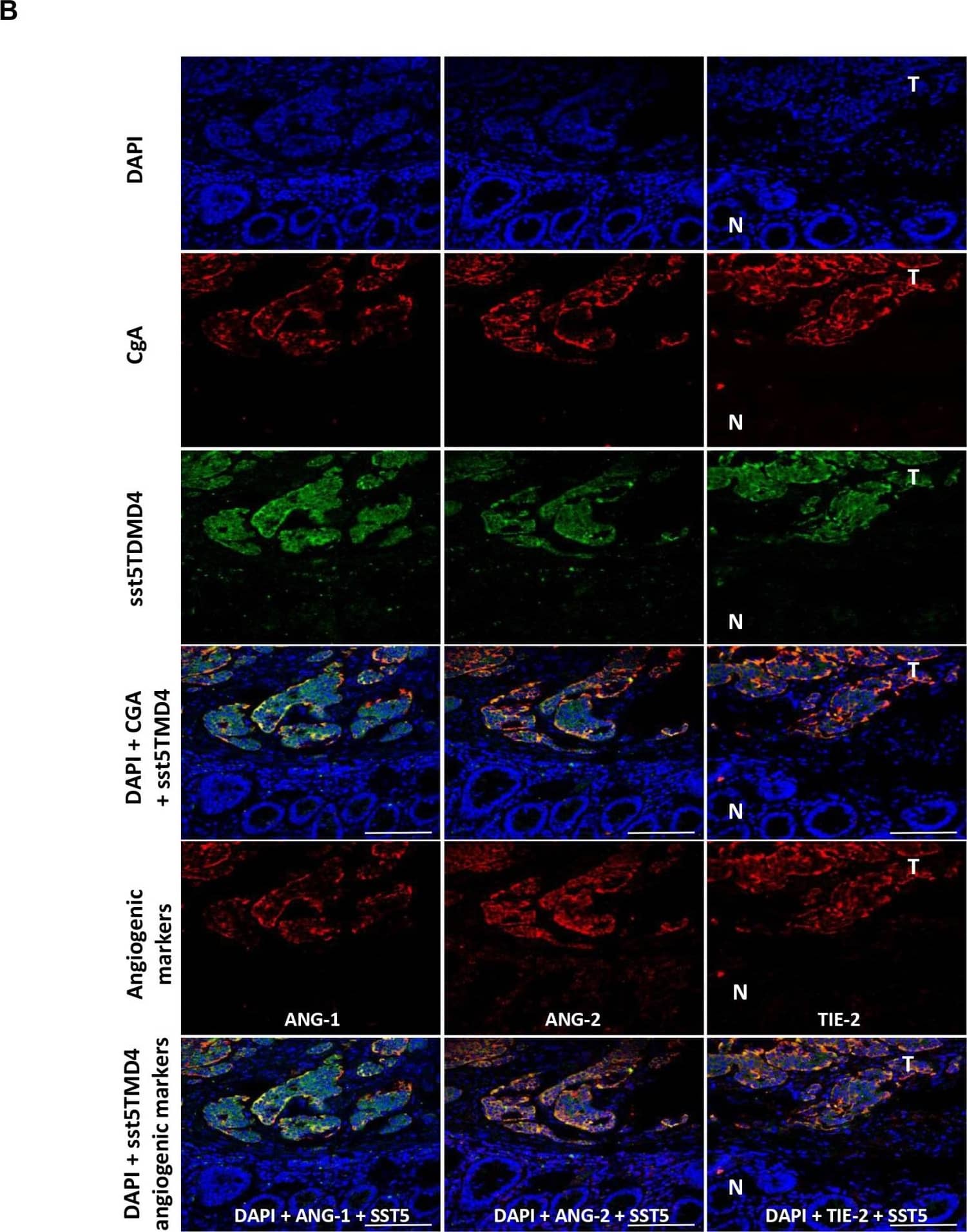
Detection of Human Angiopoietin-1 by Immunohistochemistry Expression of sst5TMD4 and co-localization with angiogenic marker in GEP-NET(A) Analysis of expression of angiogenic molecules and sst5TMD4 by specific serial immunohistochemistry in a pancreatic NET. Original magnification ×100 and ×400 (insets). N: normal tissue; T: tumor tissue. For specific immunostaining techniques see the “Materials and methods” section. (B) Expression of sst5TMD4 and angiogenic molecules by triple immunofluorescence in a gastrointestinal NET sample. Original magnification ×400. N: normal tissue; T: tumor tissue. For specific immunofluorescence techniques see the “Materials and methods” section. Scale bar for 100 μm is represented with a line for each Figure. Image collected and cropped by CiteAb from the following open publication (https://pubmed.ncbi.nlm.nih.gov/26673010), licensed under a CC-BY license. Not internally tested by R&D Systems.
Preparation and Storage
- 12 months from date of receipt, -20 to -70 °C as supplied.
- 1 month, 2 to 8 °C under sterile conditions after reconstitution.
- 6 months, -20 to -70 °C under sterile conditions after reconstitution.
Background: Angiopoietin-1
Angiopoietin-1 (Ang-1) and Angiopoietin-2 (Ang-2) are two closely related secreted ligands which bind with similar affinity to Tie-2, a receptor tyrosine kinase with immunoglobulin and epidermal growth factor homology domains expressed primarily on endothelial cells and early hematopoietic cells. Tie-2 and angiopoietins have been shown to play critical roles in embryogenic angiogenesis and in maintaining the integrity of the adult vasculature (1).
Ang-1 cDNA encodes a 498 amino acid (aa) residue precursor protein that contains a coiled-coiled domain near the amino-terminus and a fibrinogen-like domain at the C-terminus. Human Ang-1 shares approximately 97% and 60% amino acid sequence identity with mouse Ang-1 and human Ang-2, respectively (1, 2). Ang-1 activates Tie-2 signaling on endothelial cells to promote chemotaxis, cell survival, cell sprouting, vessel growth and stabilization (1, 3, 4). Ang-2 has alternatively been reported to be an antagonist for Ang-1 induced Tie-2 signaling as well as an agonist for Tie-2 signaling, depending on the cell context (5).
- Jones, N. et al. (2001) Nat. Rev. Mol. Cell Biol. 2:257.
- Davis, S. et al. (1996) Cell 87:1161.
- Witzenbichler, B. et al. (1998) J. Biol. Chem. 273:18514.
- Papapetropoulos, A. et al. (1999) Lab. Inest. 79:213.
- Teichert-Kuliszewska, K. et al. (2001) Cardiovasc. Res. 49:659.
Product Datasheets
Citations for Human/Mouse Angiopoietin-1 Antibody
R&D Systems personnel manually curate a database that contains references using R&D Systems products. The data collected includes not only links to publications in PubMed, but also provides information about sample types, species, and experimental conditions.
15
Citations: Showing 1 - 10
Filter your results:
Filter by:
-
Non-neutralizing antibodies increase endogenous circulating Ang1 levels
Authors: Zheng C, Toth J, Bigwarfe T et al.
MAbs
-
Muscle cell derived angiopoietin-1 contributes to both myogenesis and angiogenesis in the ischemic environment
Authors: Joseph M. McClung, Jessica L. Reinardy, Sarah B. Mueller, Timothy J. McCord, Christopher D. Kontos, David A. Brown et al.
Frontiers in Physiology
-
Impact of heat therapy on recovery after eccentric exercise in humans
Authors: Kyoungrae Kim, Shihuan Kuang, Qifan Song, Timothy P. Gavin, Bruno T. Roseguini
Journal of Applied Physiology
-
Pericyte Loss Leads to Capillary Stalling Through Increased Leukocyte-Endothelial Cell Interaction in the Brain
Authors: YG Choe, JH Yoon, J Joo, B Kim, SP Hong, GY Koh, DS Lee, WY Oh, Y Jeong
Frontiers in Cellular Neuroscience, 2022-03-11;16(0):848764.
Species: Mouse
Sample Types: Protein Lysates
Applications: Western Blot -
Loss of Endothelial Glycocalyx Hyaluronan Impairs Endothelial Stability and Adaptive Vascular Remodeling After Arterial Ischemia
Authors: G Wang, MR de Vries, WMPJ Sol, AM van Oevere, HC de Boer, AJ van Zonnev, PHA Quax, TJ Rabelink, BM van den Be
Cells, 2020-03-29;9(4):.
Species: Mouse
Sample Types: Whole Tissue
Applications: IHC -
EphA4/Tie2 crosstalk regulates leptomeningeal collateral remodeling following ischemic stroke
Authors: B Okyere, WA Mills Iii, X Wang, M Chen, J Chen, A Hazy, Y Qian, JB Matson, MH Theus
J. Clin. Invest., 2020-02-03;0(0):.
Species: Mouse
Sample Types: Tissue Homogenates
Applications: Western Blot -
The Fibrin Cleavage Product B?15-42 Channels Endothelial and Tubular Regeneration in the Post-acute Course During Murine Renal Ischemia Reperfusion Injury
Authors: D Fischer, C Seifen, P Baer, M Jung, C Mertens, B Scheller, K Zacharowsk, R Hofmann, TJ Maier, A Urbschat
Front Pharmacol, 2018-04-27;9(0):369.
Species: Mouse
Sample Types: Tissue Homogenates
Applications: Western Blot -
Ang-2 but not Ang-1 expression in perivascular soft tissue tumors
Authors: Swati Shrestha
J Orthop, 2016-11-28;14(1):147-153.
Species: Human
Sample Types: Whole Tissue
Applications: IHC-P -
Impact of p16 status on pro- and anti-angiogenesis factors in head and neck cancers.
Authors: Baruah P, Lee M, Wilson P, Odutoye T, Williamson P, Hyde N, Kaski J, Dumitriu I
Br J Cancer, 2015-07-14;113(4):653-9.
Species: Human
Sample Types: Whole Tissue
Applications: IHC-P -
Angiopoietin-4 promotes glioblastoma progression by enhancing tumor cell viability and angiogenesis.
Authors: Brunckhorst MK, Wang H, Lu R, Yu Q
Cancer Res., 2010-09-07;70(18):7283-93.
Species: Human
Sample Types: Cell Lysates
Applications: Western Blot -
Integrin binding angiopoietin-1 monomers reduce cardiac hypertrophy.
Authors: Dallabrida SM, Ismail NS, Pravda EA, Parodi EM, Dickie R, Durand EM, Lai J, Cassiola F, Rogers RA, Rupnick MA
FASEB J., 2008-05-23;22(8):3010-23.
Species: Mouse
Sample Types: Tissue Homogenates
Applications: Western Blot -
Angiogenesis markers (VEGF, soluble receptor of VEGF and angiopoietin-1) in very early arthritis and their association with inflammation and joint destruction.
Authors: Clavel G, Bessis N, Lemeiter D, Fardellone P, Mejjad O, Menard JF, Pouplin S, Boumier P, Vittecoq O, Le Loet X, Boissier MC
Clin. Immunol., 2007-06-08;124(2):158-64.
Species: Human
Sample Types: Serum
Applications: ELISA Development -
Enhancement of angiogenic effectors through hypoxia-inducible factor in preterm primate lung in vivo.
Authors: Asikainen TM, Waleh NS, Schneider BK, Clyman RI, White CW
Am. J. Physiol. Lung Cell Mol. Physiol., 2006-05-05;291(4):L588-95.
Species: Primate - Papio anubis (Olive Baboon)
Sample Types: Tissue Homogenates
Applications: Western Blot -
Activation of hypoxia-inducible factors in hyperoxia through prolyl 4-hydroxylase blockade in cells and explants of primate lung.
Authors: Asikainen TM, Schneider BK, Waleh NS, Clyman RI, Ho WB, Flippin LA, Gunzler V, White CW
Proc. Natl. Acad. Sci. U.S.A., 2005-07-11;102(29):10212-7.
Species: Human
Sample Types: Cell Lysates
Applications: Western Blot -
Presence of sst5TMD4, a truncated splice variant of the somatostatin receptor subtype 5, is associated to features of increased aggressiveness in pancreatic neuroendocrine tumors.
Authors: Sampedro-Nunez M, Luque RM, Ramos-Levi AM et al.
Oncotarget
FAQs
No product specific FAQs exist for this product, however you may
View all Antibody FAQsReviews for Human/Mouse Angiopoietin-1 Antibody
Average Rating: 2.5 (Based on 2 Reviews)
Have you used Human/Mouse Angiopoietin-1 Antibody?
Submit a review and receive an Amazon gift card.
$25/€18/£15/$25CAN/¥75 Yuan/¥2500 Yen for a review with an image
$10/€7/£6/$10 CAD/¥70 Yuan/¥1110 Yen for a review without an image
Filter by:

