Human M-CSF R PE-conjugated Antibody Summary
Ile20-Glu512 (Pro54Ala)
Accession # P07333.2
Applications
Please Note: Optimal dilutions should be determined by each laboratory for each application. General Protocols are available in the Technical Information section on our website.
Scientific Data
 View Larger
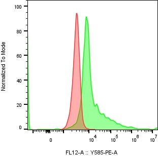
Detection of M‑CSF R in Human Blood Monocytes by Flow Cytometry. Human peripheral blood monocytes were stained with Mouse Anti-Human M-CSF R PE-conjugated Monoclonal Antibody (Catalog # FAB329P, filled histogram) or isotype control antibody (Catalog # IC002P, open histogram). View our protocol for Staining Membrane-associated Proteins.
Preparation and Storage
- 12 months from date of receipt, 2 to 8 °C as supplied.
Background: M-CSF R/CD115
M-CSF Receptor (M-CSF R), the product of the c-fms proto-oncogene, is a member of the type III subfamily of receptor tyrosine kinases that also includes receptors for SCF and PDGF. These receptors each contain five immunoglobulin-like domains in their extracellular domain (ECD) and a split kinase domain in their intracellular region (1-4). M-CSF Receptor is expressed primarily on cells of the monocyte/macrophage lineage, dendritic cells, stem cells and in the developing placenta (1). Human M-CSF Receptor cDNA encodes a 972 amino acid (aa) type I membrane protein with a 19 aa signal peptide, a 493 aa extracellular region containing the ligand-binding domain, a 25 aa transmembrane domain, and a 435 aa cytoplasmic domain. The human M-CSF R ECD shares 60%, 64%, 72%, 75%, 75%, and 76% aa identity with mouse, rat, bovine, canine, feline, and equine M-CSF R, respectively. Activators of protein kinase C induce TACE/ADAM17 cleavage of the M-CSF Receptor, releasing the functional ligand-binding extracellular domain (5). M-CSF binding induces receptor homodimerization, resulting in transphosphorylation of specific cytoplasmic tyrosine residues and signal transduction (6). The intracellular domain of activated M-CSF R binds more than 150 proteins that affect cell proliferation, survival, differentiation and cytoskeletal reorganization. Among these, PI3Kinase, P42/44 ERK, and c-Cbl are key transducers of M-CSF R signals (3, 4). M-CSF R engagement is continuously required for macrophage survival and regulates lineage decisions and maturation of monocytes, macrophages, osteoclasts and DC (3, 4). M-CSF R and Integrin alpha v beta 3 share signaling pathways during osteoclastogenesis and deletion of either causes osteopetrosis (7, 8). In the brain, microglia expressing increased M-CSF R are concentrated with Alzheimers a beta peptide, but their role in pathogenesis is unclear (9, 10).
- deParseval, N. et al. (1993) Nucleic Acids Res. 21:750.
- Rothwell, V.M. and L.R. Rohrschneider (1987) Oncogene Res. 1:311.
- Chitu, V. and E.R. Stanley (2006) Curr. Opin. Immunol. 18:39.
- Ross, F.P. and S.L. Teitelbaum (2005) Immunol. Rev. 208:88.
- Rovida, E. et al. (2001) J. Immunol. 166:1583.
- Yeung, Y. et al. (1998) J. Biol. Chem. 273:17128.
- Dai, X. et al. (2002) Blood 99:111.
- Faccio, R. et al. (2003) J. Clin. Invest. 111:749.
- Li, M. et al. (2004) J. Neurochem. 91:623.
- Mitrasinovic, O.M. et al. (2005) J. Neurosci. 25:4442.
Product Datasheets
Citations for Human M-CSF R PE-conjugated Antibody
R&D Systems personnel manually curate a database that contains references using R&D Systems products. The data collected includes not only links to publications in PubMed, but also provides information about sample types, species, and experimental conditions.
5
Citations: Showing 1 - 5
Filter your results:
Filter by:
-
The Hematopoietic Differentiation and Production of Mature Myeloid Cells from Human Pluripotent Stem Cells
Authors: Kyung-Dal Choi, Maxim Vodyanik, Igor I. Slukvin
Nature Protocols
-
Langerin-expressing dendritic cells in human tissues are related to CD1c+ dendritic cells and distinct from Langerhans cells and CD141high XCR1+ dendritic cells.
Authors: Bigley V, McGovern N, Milne P, Dickinson R, Pagan S, Cookson S, Haniffa M, Collin M
J Leukoc Biol, 2014-12-16;97(4):627-34.
Species: Human
Sample Types: Whole Cells
Applications: Flow Cytometry -
Phenotyping of human melanoma cells reveals a unique composition of receptor targets and a subpopulation co-expressing ErbB4, EPO-R and NGF-R.
Authors: Mirkina I, Hadzijusufovic E, Krepler C, Mikula M, Mechtcheriakova D, Strommer S, Stella A, Jensen-Jarolim E, Holler C, Wacheck V, Pehamberger H, Valent P
PLoS ONE, 2014-01-29;9(1):e84417.
Species: Human
Sample Types: Whole Cells
Applications: Flow Cytometry -
Systematic cytokine receptor profiling reveals GM-CSF as a novel TLR-independent activator of human plasmacytoid predendritic cells.
Authors: Ghirelli C, Zollinger R, Soumelis V
Blood, 2010-04-09;115(24):5037-40.
Species: Human
Sample Types: Whole Cells
Applications: Flow Cytometry -
Inhibition of RANK expression and osteoclastogenesis by TLRs and IFN-gamma in human osteoclast precursors.
Authors: Ji JD, Park-Min KH, Shen Z, Fajardo RJ, Goldring SR, McHugh KP, Ivashkiv LB
J. Immunol., 2009-11-04;183(11):7223-33.
Species: Human
Sample Types: Whole Cells
Applications: Flow Cytometry
FAQs
No product specific FAQs exist for this product, however you may
View all Antibody FAQsReviews for Human M-CSF R PE-conjugated Antibody
Average Rating: 4 (Based on 1 Review)
Have you used Human M-CSF R PE-conjugated Antibody?
Submit a review and receive an Amazon gift card.
$25/€18/£15/$25CAN/¥75 Yuan/¥2500 Yen for a review with an image
$10/€7/£6/$10 CAD/¥70 Yuan/¥1110 Yen for a review without an image
Filter by:


