Human IL-3 Antibody Summary
Ala20-Phe152
Accession # Q6GS87
Applications
Please Note: Optimal dilutions should be determined by each laboratory for each application. General Protocols are available in the Technical Information section on our website.
Scientific Data
 View Larger
Cell Proliferation Induced by IL‑3 and Neutralization by Human IL‑3 Antibody. Recombinant Human IL‑3 (Catalog # 203-IL) stimulates proliferation in the TF‑1 human erythroleukemic cell line in a dose-dependent manner (orange line). Proliferation elicited by Recombinant Human IL‑3 (1.25 ng/mL) is neutralized (green line) by increasing concentrations of Mouse Anti-Human IL‑3 Monoclonal Antibody (Catalog # MAB203). The ND50 is typically 0.03-0.08 µg/mL.
 View Larger
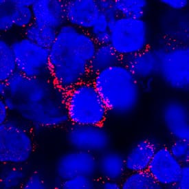
IL‑3 in Human PBMCs. IL-3 was detected in immersion fixed human peripheral blood mononuclear cells (PBMCs) using Mouse Anti-Human IL-3 Monoclonal Antibody (Catalog # MAB203) at 25 µg/mL for 3 hours at room temperature. Cells were stained using the NorthernLights™ 557-conjugated Anti-Mouse IgG Secondary Antibody (red; Catalog # NL007) and counterstained with DAPI (blue). Specific staining was localized to cytoplasm. View our protocol for Fluorescent ICC Staining of Cells on Coverslips.
Preparation and Storage
- 12 months from date of receipt, -20 to -70 °C as supplied.
- 1 month, 2 to 8 °C under sterile conditions after reconstitution.
- 6 months, -20 to -70 °C under sterile conditions after reconstitution.
Background: IL-3
Interleukin 3 is a pleiotropic factor produced primarily by activated T cells that can stimulate the proliferation and differentiation of pluripotent hematopoietic stem cells as well as various lineage committed progenitors. In addition, IL-3 also affects the functional activity of mature mast cells, basophils, eosinophils and macrophages. Because of its multiple functions and targets, it was originally studied under different names, including mast cell growth factor, P-cell stimulating factor, burst promoting activity, multi-colony stimulating factor, thy-1 inducing factor and WEHI-3 growth factor. In addition to activated T cells, other cell types such as human thymic epithelial cells, activated murine mast cells, murine keratinocytes and neurons/astrocytes can also produce IL-3. At the amino acid sequence level, mature human and murine IL-3 share only 29% sequence identity. Consistent with this lack of homology, IL-3 activity is highly species-specific and human IL-3 does not show activity on murine cells.
IL-3 exerts its biological activities through binding to specific cell surface receptors. The high affinity receptor responsible for IL-3 signaling is composed of at least two subunits, an IL-3 specific alpha chain which binds IL-3 with low affinity and a common beta chain that is shared by the IL-5 and GM-CSF high-affinity receptors. Although the beta chain itself does not bind IL-3, it confers high-affinity IL-3 binding in the presence of the alpha chain. Receptors for IL-3 are present on bone marrow progenitors, macrophages, mast cells, eosinophils, megakaryocytes, basophils and various myeloid leukemic cells.
Product Datasheets
Citations for Human IL-3 Antibody
R&D Systems personnel manually curate a database that contains references using R&D Systems products. The data collected includes not only links to publications in PubMed, but also provides information about sample types, species, and experimental conditions.
4
Citations: Showing 1 - 4
Filter your results:
Filter by:
-
Hematopoietic stem cell cytokines and fibroblast growth factor-2 stimulate human endothelial cell-pericyte tube co-assembly in 3D fibrin matrices under serum-free defined conditions.
Authors: Smith A, Bowers S, Stratman A, Davis G
PLoS ONE, 2013-12-31;8(12):e85147.
Species: Human
Sample Types: Whole Cells
Applications: Neutralization -
Anti-GM-CSF autoantibodies in patients with cryptococcal meningitis.
Authors: Rosen L, Freeman A, Yang L, Jutivorakool K, Olivier K, Angkasekwinai N, Suputtamongkol Y, Bennett J, Pyrgos V, Williamson P, Ding L, Holland S, Browne S
J Immunol, 2013-03-18;190(8):3959-66.
Species: Human
Sample Types: Plasma
Applications: Flow Cytometry -
Expression of functional interleukin-3 receptors on Hodgkin and Reed-Sternberg cells.
Authors: Aldinucci D, 2019, Poletto D, Gloghini A, Nanni P, Degan M, Perin T, Ceolin P, Rossi FM, Gattei V, Carbone A, Pinto A
439, 2002-02-01;160(2):585-96.
Species: Human
Sample Types: Whole Cells
Applications: Neutralization -
Human T-cell leukemia virus type 2 induces survival and proliferation of CD34(+) TF-1 cells through activation of STAT1 and STAT5 by secretion of interferon-gamma and granulocyte macrophage-colony-stimulating factor.
Authors: Bovolenta C, Pilotti E, Mauri M, Turci M, Ciancianaini P, Fisicaro P, Bertazzoni U, Poli G, Casoli C
Blood, 2002-01-01;99(1):224-31.
Species: Human
Sample Types: Whole Cells
Applications: Neutralization
FAQs
-
Does Human IL-3 Antibody, Catalog # MAB203, Clone # 4806, have kappa or lambda light chain?
MAB203, Clone # 4806 has kappa light chain.
Reviews for Human IL-3 Antibody
There are currently no reviews for this product. Be the first to review Human IL-3 Antibody and earn rewards!
Have you used Human IL-3 Antibody?
Submit a review and receive an Amazon gift card.
$25/€18/£15/$25CAN/¥75 Yuan/¥2500 Yen for a review with an image
$10/€7/£6/$10 CAD/¥70 Yuan/¥1110 Yen for a review without an image


