Human IL-17/IL-17A Antibody Summary
Ile20-Ala155
Accession # Q16552
Applications
Please Note: Optimal dilutions should be determined by each laboratory for each application. General Protocols are available in the Technical Information section on our website.
Scientific Data
 View Larger
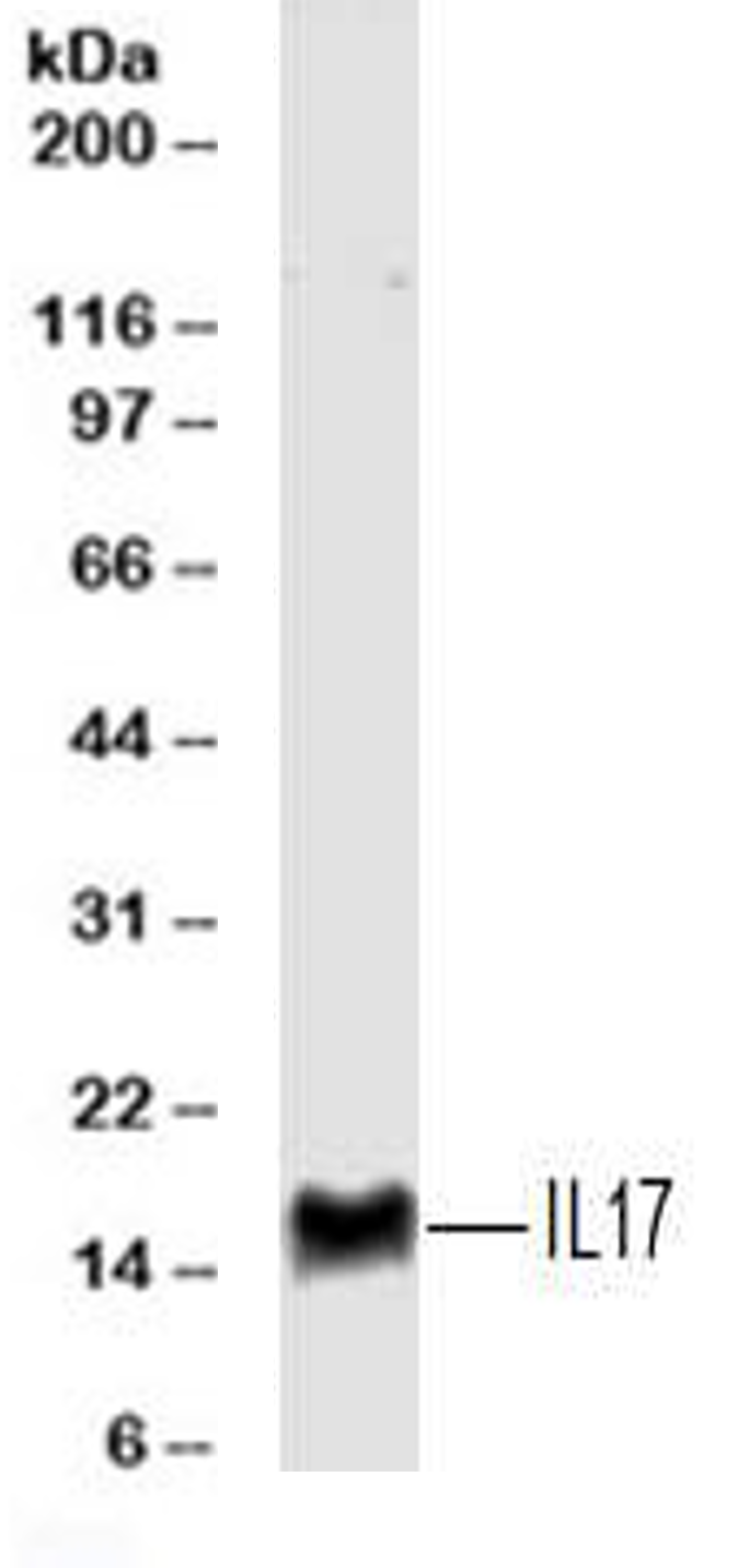
Detection of IL-17/IL-17A Mouse by Western Blot. Western blot shows lysates of purified naïve human CD4+T cells orin vitrodifferentiated human Th17 CD4+ T cells. PVDF membrane was probed with 2 µg/mL of Mouse Anti-Human IL-17 Monoclonal Antibody (Catalog # MAB3171) followed by HRP-conjugated Anti-Mouse IgG Secondary Antibody (Catalog # HAF007). A specific band was detected for IL-17 at approximately 17 kDa (as indicated). This experiment was conducted under reducing conditions and using Immunoblot Buffer Group 1.
 View Larger
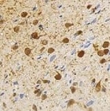
IL‑17/IL‑17A in Human Crohn's Disease Intestine. IL-17/IL-17A was detected in immersion fixed paraffin-embedded sections of human Crohn's disease intestine using Mouse Anti-Human IL-17/IL-17A Monoclonal Antibody (Catalog # MAB3171) at 0.5 µg/mL for 1 hour at room temperature followed by incubation with the Anti-Mouse IgG VisUCyte™ HRP Polymer Antibody (Catalog # VC001). Before incubation with the primary antibody, tissue was subjected to heat-induced epitope retrieval using Antigen Retrieval Reagent-Basic (Catalog # CTS013). Tissue was stained using DAB (brown) and counterstained with hematoxylin (blue). Specific staining was localized to lymphocytes. View our protocol for IHC Staining with VisUCyte HRP Polymer Detection Reagents.
 View Larger
Immunoprecipitation of Human IL-17. Human IL-17 was immunoprecipitated from 100 µg of humanin vitrodifferentiated Th17 cell lysate following incubation with 3 µg Mouse Anti-Human IL-17 Monoclonal Antibody (Catalog # MAB3171) or isotype control antibody (Catalog # MAB002) overnight at 4 °C. IL-17-antibody complexes were absorbed using anti-mouse agarose beads. Immunoprecipitated IL-17 was detected by Western blot using 1 µg/mL Goat Anti-Human IL-17 Antigen Affinity-purified Polyclonal Antibody (Catalog # AF-317-NA). View our recommended buffer recipes for immunoprecipitation.
 View Larger
Detection of IL‑17 in Human PBMCs by Flow Cytometry. Human peripheral blood mononuclear cells were unstimulated (light orange filled histogram) or treated with 50 ng/mL PMA and 250 ng/mL Ca2+ionomycin for 16 hours, then stained with Mouse Anti-Human IL-17 Monoclonal Antibody (Catalog # MAB3171, dark orange filled histogram) or isotype control antibody (Catalog # MAB002, open histogram), followed by Allophycocyanin-conjugated Anti-Mouse IgG F(ab')2Secondary Antibody (Catalog # F0101B). To facilitate intracellular staining, cells were fixed with paraformaldehyde and permeabilized with saponin.
Preparation and Storage
- 12 months from date of receipt, -20 to -70 °C as supplied.
- 1 month, 2 to 8 °C under sterile conditions after reconstitution.
- 6 months, -20 to -70 °C under sterile conditions after reconstitution.
Background: IL-17/IL-17A
Interleukin-17 (IL-17) is a pro-inflammatory cytokine secreted by activated T cells. It is the prototype member of the IL-17 family that also includes IL-17B, C, D, E, and F.
Product Datasheets
Citations for Human IL-17/IL-17A Antibody
R&D Systems personnel manually curate a database that contains references using R&D Systems products. The data collected includes not only links to publications in PubMed, but also provides information about sample types, species, and experimental conditions.
10
Citations: Showing 1 - 10
Filter your results:
Filter by:
-
Clarithromycin Enhances the Antibacterial Activity and Wound Healing Capacity in Type 2 Diabetes Mellitus by Increasing LL-37 Load on Neutrophil Extracellular Traps
Authors: Athanasios Arampatzioglou, Dimitrios Papazoglou, Theocharis Konstantinidis, Akrivi Chrysanthopoulou, Alexandros Mitsios, Iliana Angelidou et al.
Frontiers in Immunology
-
IL-17 Induces Autophagy Dysfunction to Promote Inflammatory Cell Death and Fibrosis in Keloid Fibroblasts via the STAT3 and HIF-1 alpha Dependent Signaling Pathways
Authors: Seon-Yeong Lee, A Ram Lee, Jeong Won Choi, Chae Rim Lee, Keun-Hyung Cho, Jung Ho Lee et al.
Frontiers in Immunology
-
Impaired IL-17 Signaling Pathway Contributes to the Increased Collagen Expression in Scleroderma Fibroblasts
Authors: Taiji Nakashima, Masatoshi Jinnin, Keitaro Yamane, Noritoshi Honda, Ikko Kajihara, Takamitsu Makino et al.
The Journal of Immunology
-
Establishment of a humanized mouse model of keloid diseases following the migration of patient immune cells to the lesion: Patient-derived keloid xenograft (PDKX) model
Authors: Lee, AR;Lee, SY;Choi, JW;Um, IG;Na, HS;Lee, JH;Cho, ML;
Experimental & molecular medicine
Applications: IHC, Confocal Imaging -
Establishment of a humanized mouse model of keloid diseases following the migration of patient immune cells to the lesion: Patient-derived keloid xenograft (PDKX) model
Authors: Lee, AR;Lee, SY;Choi, JW;Um, IG;Na, HS;Lee, JH;Cho, ML;
Experimental & molecular medicine
Species: Mouse
Sample Types: Whole Tissue
Applications: IHC -
Impact of Adalimumab Treatment on Interleukin-17 and Interleukin-17 Receptor Expression in Skin and Synovium of Psoriatic Arthritis Patients with Mild Psoriasis
Authors: JW Bolt, AW van Kuijk, MBM Teunissen, D van der Co, S Aarrass, DM Gerlag, PP Tak, MG van de San, MC Lebre, LGM van Baarse
Biomedicines, 2022-01-29;10(2):.
Species: Human
Sample Types: Whole Tissue
Applications: IHC -
Comparison of three different ELISAs for the detection of recombinant, native and plasma IL-17A
Authors: MB Ismail, SO Åkefeldt, M Lourda, D Gavhed, R Gayet, M Aricò, JI Henter, C Delprat, H Valentin
MethodsX, 2020-07-16;7(0):100997.
Species: Human
Sample Types: Plasma, Recombinant Protein
Applications: ELISA Capture -
Patients with cystic fibrosis have inducible IL-17+IL-22+ memory cells in lung draining lymph nodes.
Authors: Chan Y, Chen K, Duncan S, Lathrop K, Latoche J, Logar A, Pociask D, Wahlberg B, Ray P, Ray A, Pilewski J, Kolls J
J Allergy Clin Immunol, 2012-07-11;131(4):1117-29, 1129.
Species: Human
Sample Types: Whole Tissue
Applications: IHC -
Langerhans cell histiocytosis reveals a new IL-17A-dependent pathway of dendritic cell fusion.
Authors: Coury F, Annels N, Rivollier A, Olsson S, Santoro A, Speziani C, Azocar O, Flacher M, Djebali S, Tebib J, Brytting M, Egeler RM, Rabourdin-Combe C, Henter JI, Arico M, Delprat C
Nat. Med., 2007-12-23;14(1):81-7.
Species: Human
Sample Types: Whole Tissue
Applications: IHC-P -
Angiogenic Role of Mesothelium-Derived Chemokine CXCL1 During Unfavorable Peritoneal Tissue Remodeling in Patients Receiving Peritoneal Dialysis as Renal Replacement Therapy
Authors: Rusan Ali Catar, Maria Bartosova, Edyta Kawka, Lei Chen, Iva Marinovic, Conghui Zhang et al.
Frontiers in Immunology
FAQs
No product specific FAQs exist for this product, however you may
View all Antibody FAQsReviews for Human IL-17/IL-17A Antibody
Average Rating: 4.3 (Based on 3 Reviews)
Have you used Human IL-17/IL-17A Antibody?
Submit a review and receive an Amazon gift card.
$25/€18/£15/$25CAN/¥75 Yuan/¥2500 Yen for a review with an image
$10/€7/£6/$10 CAD/¥70 Yuan/¥1110 Yen for a review without an image
Filter by:


