Human IgM Antibody Summary
*Small pack size (-SP) is supplied either lyophilized or as a 0.2 µm filtered solution in PBS.
Applications
This antibody functions as an ELISA detection antibody when paired with Rabbit Anti-Human IgM Monoclonal Antibody (Catalog # MAB9435).
This product is intended for assay development on various assay platforms requiring antibody pairs.
Please Note: Optimal dilutions should be determined by each laboratory for each application. General Protocols are available in the Technical Information section on our website.
Scientific Data
 View Larger
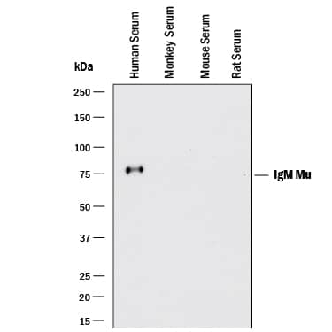
Detection of Human IgM by Western Blot. Western blot shows lysates of human serum, monkey serum (negative control), mouse serum (negative control), and rat serum (negative control). PVDF membrane was probed with 0.1 µg/mL of Rabbit Anti-Human IgM Monoclonal Antibody (Catalog # MAB94351) followed by HRP-conjugated Anti-Rabbit IgG Secondary Antibody (HAF008). A specific band was detected for IgM at approximately 75 kDa (as indicated). This experiment was conducted under reducing conditions and using Immunoblot Buffer Group 1.
 View Larger
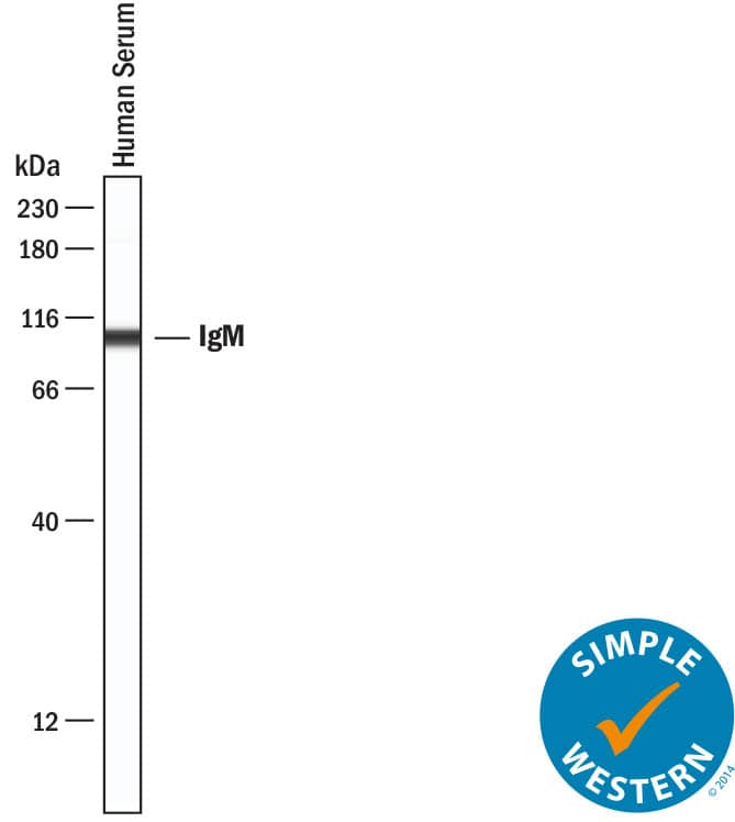
Detection of Human IgM by Simple WesternTM. Simple Western lane view shows lysates of human serum, loaded at 1:4000 dilution. A specific band was detected for IgM at approximately 103 kDa (as indicated) using 5 µg/mL of Rabbit Anti-Human IgM Monoclonal Antibody (Catalog # MAB94351). This experiment was conducted under reducing conditions and using the 12‑230 kDa separation system.
 View Larger
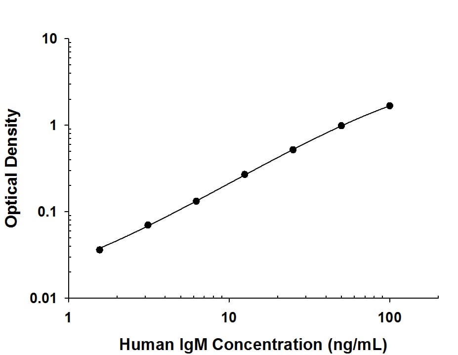
Human IgM ELISA Standard Curve. Recombinant Human IgM protein was serially diluted 2-fold and captured by Rabbit Anti-Human IgM Monoclonal Antibody (MAB9435) coated on a Clear Polystyrene Microplate (DY990). Rabbit Anti-Human IgM Monoclonal Antibody (Catalog # MAB94351) was biotinylated and incubated with the protein captured on the plate. Detection of the standard curve was achieved by incubating Streptavidin-HRP (DY998) followed by Substrate Solution (DY999) and stopping the enzymatic reaction with Stop Solution (DY994).
 View Larger
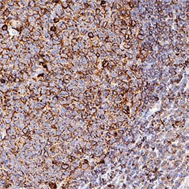
Detection of IgM in Human Tonsil. IgM was detected in immersion fixed paraffin-embedded sections of human tonsil using Rabbit Anti-Human IgM Monoclonal Antibody (Catalog # MAB94351) at 3 µg/ml for 1 hour at room temperature followed by incubation with the HRP-conjugated Anti-Rabbit IgG Secondary Antibody (Catalog # HAF008) or the Anti-Rabbit IgG VisUCyte™ HRP Polymer Antibody (Catalog # VC003). Before incubation with the primary antibody, tissue was subjected to heat-induced epitope retrieval using VisUCyte Antigen Retrieval Reagent-Basic (Catalog # VCTS021). Tissue was stained using DAB (brown) and counterstained with hematoxylin (blue). Specific staining was localized to the membrane of B cells. View our protocol for Chromogenic IHC Staining of Paraffin-embedded Tissue Sections.
Preparation and Storage
- 12 months from date of receipt, -20 to -70 °C as supplied.
- 1 month, 2 to 8 °C under sterile conditions after reconstitution.
- 6 months, -20 to -70 °C under sterile conditions after reconstitution.
Background: IgM
R&D Systems offers a range of secondary antibodies and controls for flow cytometry, immunohistochemistry, and Western blotting. We provide species-specific secondary antibodies that are available with a variety of conjugated labels.
Our NorthernLights fluorescent secondary antibodies are bright and resistant to photobleaching. We are currently offering secondary antibodies recognizing mouse, rat, goat, sheep, and rabbit IgG as well as chicken IgY. These reagents are available with three distinct excitation and emission maxima, making them ideal for multi-color fluorescence microscopy.
Product Datasheets
FAQs
No product specific FAQs exist for this product, however you may
View all Antibody FAQsReviews for Human IgM Antibody
There are currently no reviews for this product. Be the first to review Human IgM Antibody and earn rewards!
Have you used Human IgM Antibody?
Submit a review and receive an Amazon gift card.
$25/€18/£15/$25CAN/¥75 Yuan/¥2500 Yen for a review with an image
$10/€7/£6/$10 CAD/¥70 Yuan/¥1110 Yen for a review without an image
