Human HGFR/c-MET PE-conjugated Antibody Summary
Glu25-Thr932
Accession # P08581
Applications
Please Note: Optimal dilutions should be determined by each laboratory for each application. General Protocols are available in the Technical Information section on our website.
Scientific Data
 View Larger
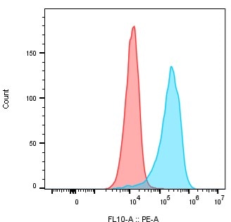
Detection of HGF R/c‑MET in MDA‑MB‑231 Human Cell Line by Flow Cytometry. MDA-MB-231 human breast cancer cell line was stained with Mouse Anti-Human HGF R/c-MET PE-conjugated Monoclonal Antibody (Catalog # FAB3582P, filled histogram) or isotype control antibody (Catalog # IC002P, open histogram). View our protocol for Staining Membrane-associated Proteins.
Preparation and Storage
- 12 months from date of receipt, 2 to 8 °C as supplied.
Background: HGFR/c-MET
HGF R, also known as Met (from N-methyl-N’-nitro-N-nitrosoguanidine induced), is a glycosylated receptor tyrosine kinase that plays a central role in epithelial morphogenesis and cancer development. HGF R is synthesized as a single chain precursor which undergoes cotranslational proteolytic cleavage. This generates a mature HGF R that is a disulfide-linked dimer composed of a 50 kDa extracellular alpha chain and a 145 kDa transmembrane beta chain (1, 2). The extracellular domain (ECD) contains a seven bladed beta -propeller sema domain, a cysteine-rich PSI/MRS, and four Ig-like E-set domains, while the cytoplasmic region includes the tyrosine kinase domain (3, 4). Proteolysis and alternate splicing generate additional forms of human HGF R which either lack of the kinase domain, consist of secreted extracellular domains, or are deficient in proteolytic separation of the alpha and beta chains (5-7). The sema domain, which is formed by both the alpha and beta chains of HGF R, mediates both ligand binding and receptor dimerization (3, 8). Ligand-induced tyrosine phosphorylation in the cytoplasmic region activates the kinase domain and provides docking sites for multiple SH2-containing molecules (9, 10). HGF stimulation induces HGF R downregulation via internalization and proteasome-dependent degradation (11). In the absence of ligand, HGF R forms non-covalent complexes with a variety of membrane proteins including CD44v6, CD151, EGF R, Fas, Integrin alpha 6/ beta 4, Plexins B1, 2, 3, and MSP R/Ron (12-19). Ligation of one complex component triggers activation of the other, followed by cooperative signaling effects (12-19). Formation of some of these heteromeric complexes is a requirement for epithelial cell morphogenesis and tumor cell invasion (12, 16, 17). Paracrine induction of epithelial cell scattering and branching tubulogenesis results from the stimulation of HGF R on undifferentiated epithelium by HGF released from neighboring mesenchymal cells (20). Genetic polymorphisms, chromosomal translocation, over-expression, and additional splicing and proteolytic cleavage of HGF R have been described in a wide range of cancers (1). Within the ECD, human HGF R shares 86-88% amino acid sequence identity with canine, mouse, and rat HGF R.
- Birchmeier, C. et al. (2003) Nat. Rev. Mol. Cell Biol. 4:915.
- Corso, S. et al. (2005) Trends Mol. Med. 11:284.
- Gherardi, E. et al. (2003) Proc. Natl. Acad. Sci. USA 100:12039.
- Park, M. et al. (1987) Proc. Natl. Acad. Sci. USA 84:6379.
- Crepaldi, T. et al. (1994) J. Biol. Chem. 269:1750.
- Prat, M. et al. (1991) Mol. Cell. Biol. 12:5954.
- Rodrigues, G.A. et al. (1991) Mol. Cell. Biol. 11:2962.
- Kong-Beltran, M. et al. (2004) Cancer Cell 6:75.
- Naldini, L. et al. (1991) Mol. Cell. Biol. 11:1793.
- Ponzetto, C. et al. (1994) Cell 77:261.
- Jeffers, M. et al. (1997) Mol. Cell. Biol. 17:799.
- Orian-Rousseau, V. et al. (2002) Genes Dev. 16:3074.
- Klosek, S.K. et al. (2005) Biochem. Biophys. Res. Commun. 336:408.
- Jo, M. et al. (2000) J. Biol. Chem. 275:8806.
- Wang, X. et al. (2002) Mol. Cell 9:411.
- Trusolino, L. et al. (2001) Cell 107:643.
- Giordano, S. et al. (2002) Nat. Cell Biol. 4:720.
- Conrotto, P. et al. (2004) Oncogene 23:5131.
- Follenzi, A. et al. (2000) Oncogene 19:3041.
- Sonnenberg, E. et al. (1993) J. Cell Biol. 123:223.
Product Datasheets
Citations for Human HGFR/c-MET PE-conjugated Antibody
R&D Systems personnel manually curate a database that contains references using R&D Systems products. The data collected includes not only links to publications in PubMed, but also provides information about sample types, species, and experimental conditions.
11
Citations: Showing 1 - 10
Filter your results:
Filter by:
-
Coexisting cancer stem cells with heterogeneous gene amplifications, transcriptional profiles, and malignancy are isolated from single glioblastomas
Authors: De Bacco, F;Orzan, F;Crisafulli, G;Prelli, M;Isella, C;Casanova, E;Albano, R;Reato, G;Erriquez, J;D'Ambrosio, A;Panero, M;Dall'Aglio, C;Casorzo, L;Cominelli, M;Pagani, F;Melcarne, A;Zeppa, P;Altieri, R;Morra, I;Cassoni, P;Garbossa, D;Cassisa, A;Bartolini, A;Pellegatta, S;Comoglio, PM;Finocchiaro, G;Poliani, PL;Boccaccio, C;
Cell reports
Species: Human
Sample Types: Whole Cells
Applications: Flow Cytometry -
Endosomal LC3C-pathway selectively targets plasma membrane cargo for autophagic degradation
Authors: PP Coelho, GG Hesketh, A Pedersen, E Kuzmin, AN Fortier, ES Bell, CDH Ratcliffe, AC Gingras, M Park
Nature Communications, 2022-07-02;13(1):3812.
Species: Human
Sample Types: Whole Cells
Applications: Flow Cytometry -
HGF-Induced PD-L1 Expression in Head and Neck Cancer: Preclinical and Clinical Findings
Authors: V Boschert, J Teusch, A Aljasem, P Schmucker, N Klenk, A Straub, M Bittrich, A Seher, C Linz, UDA Müller-Ric, S Hartmann
Int J Mol Sci, 2020-11-20;21(22):.
Species: Human
Sample Types: Cell Lysates
Applications: Western Blot -
Efficient blockade of locally reciprocated tumor-macrophage signaling using a TAM-avid nanotherapy
Authors: SJ Wang, R Li, TSC Ng, G Luthria, MJ Oudin, M Prytyskach, RH Kohler, R Weissleder, DA Lauffenbur, MA Miller
Sci Adv, 2020-05-22;6(21):eaaz8521.
Species: Human
Sample Types: Whole Cells
Applications: Flow Cytometry -
Combination therapy with c-met inhibitor and TRAIL enhances apoptosis in dedifferentiated liposarcoma patient-derived cells
Authors: EB Jo, YS Lee, H Lee, JB Park, H Park, YL Choi, D Hong, SJ Kim
BMC Cancer, 2019-05-24;19(1):496.
Species: Human
Sample Types: Whole Cells
Applications: Flow Cytometry -
Ezrin promotes stem cell properties in pancreatic ductal adenocarcinoma
Authors: VR Penchev, YT Chang, A Begum, T Ewachiw, C Gocke, J Li, RH McMillan, Q Wang, R Anders, L Marchionni, A Maitra, A Uren, Z Rasheed, W Matsui
Mol. Cancer Res., 2019-01-17;0(0):.
Species: Human
Sample Types: Whole Cells
Applications: Flow Cytometry -
An essential receptor for adeno-associated virus infection.
Authors: Pillay S, Meyer N, Puschnik A, Davulcu O, Diep J, Ishikawa Y, Jae L, Wosen J, Nagamine C, Chapman M, Carette J
Nature, 2016-01-27;530(7588):108-12.
Species: Human
Sample Types: Whole Cells
Applications: Flow Cytometry -
Impact of Cell-surface Antigen Expression on Target Engagement and Function of an Epidermal Growth Factor Receptor x c-MET Bispecific Antibody.
Authors: Jarantow S, Bushey B, Pardinas J, Boakye K, Lacy E, Sanders R, Sepulveda M, Moores S, Chiu M
J Biol Chem, 2015-08-10;290(41):24689-704.
Species: Human
Sample Types: Whole Cells
Applications: Flow Cytometry -
Deregulated hepsin protease activity confers oncogenicity by concomitantly augmenting HGF/MET signalling and disrupting epithelial cohesion.
Authors: Tervonen T, Belitskin D, Pant S, Englund J, Marques E, Ala-Hongisto H, Nevalaita L, Sihto H, Heikkila P, Leidenius M, Hewitson K, Ramachandra M, Moilanen A, Joensuu H, Kovanen P, Poso A, Klefstrom J
Oncogene, 2015-07-13;0(0):ePub.
Species: Human
Sample Types: Whole Cells
Applications: Flow Cytometry -
Three-dimensional lung tumor microenvironment modulates therapeutic compound responsiveness in vitro--implication for drug development.
Authors: Ekert, Jason E, Johnson, Kjell, Strake, Brandy, Pardinas, Jose, Jarantow, Stephen, Perkinson, Robert, Colter, David C
PLoS ONE, 2014-03-17;9(3):e92248.
Species: Human
Sample Types: Whole Cells
Applications: Flow Cytometry -
Phenotyping of human melanoma cells reveals a unique composition of receptor targets and a subpopulation co-expressing ErbB4, EPO-R and NGF-R.
Authors: Mirkina I, Hadzijusufovic E, Krepler C, Mikula M, Mechtcheriakova D, Strommer S, Stella A, Jensen-Jarolim E, Holler C, Wacheck V, Pehamberger H, Valent P
PLoS ONE, 2014-01-29;9(1):e84417.
Species: Human
Sample Types: Whole Cells
Applications: Flow Cytometry
FAQs
No product specific FAQs exist for this product, however you may
View all Antibody FAQsReviews for Human HGFR/c-MET PE-conjugated Antibody
Average Rating: 5 (Based on 1 Review)
Have you used Human HGFR/c-MET PE-conjugated Antibody?
Submit a review and receive an Amazon gift card.
$25/€18/£15/$25CAN/¥75 Yuan/¥2500 Yen for a review with an image
$10/€7/£6/$10 CAD/¥70 Yuan/¥1110 Yen for a review without an image
Filter by:


