Human HGFR/c-MET APC-conjugated Antibody Summary
Glu25-Thr932
Accession # P08581
Applications
Please Note: Optimal dilutions should be determined by each laboratory for each application. General Protocols are available in the Technical Information section on our website.
Scientific Data
 View Larger
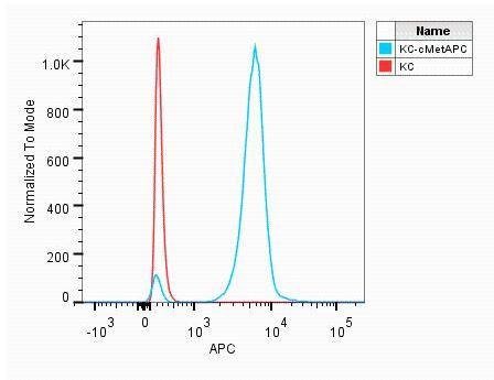
Detection of HGF R/c‑MET in MDA‑MB‑231 Human Cell Line by Flow Cytometry. MDA-MB-231 human breast cancer cell line was stained with Mouse Anti-Human HGF R/c-MET APC-conjugated Monoclonal Antibody (Catalog # FAB3582A, filled histogram) or isotype control antibody (Catalog # IC002A, open histogram). View our protocol for Staining Membrane-associated Proteins.
 View Larger
Detection of Human HGFR/c-MET by Flow Cytometry Influence of selected cytokines on MSC proliferation; receptor expression profiles and concentration. (A) MSC were seeded in 5% pHPL, tPRP and FBS, respectively, and stimulated with for 3 days with 50 ng/ml HGF, 10 ng/ml IGF-1 or 25 ng/ml bFGF. Cell counts were acquired with the CellTiter-Glo assay and then normalized to the unstimulated control to derive relative cell count values. (B)% positivity of IGF, FGF and HGF receptor expression of BM- and LA-MSC (donors 1–3, respectively) in pHPL, tPRP and FBS assessed by flow cytometry. (C – E): IGF, FGF and HGF concentrations were determined by ELISA in pHPL and tPRP supplemented medium (medium, 6 different batches); medium stored for 24 h (control medium) and conditioned by MSC (CM) (each n = 3). Symbols indicate statistically significant diffences between: * stimulation; + supplements; # MSC sources; (one symbol p < 0.05; two symbols p < 0.01). Image collected and cropped by CiteAb from the following publication (https://bmcmolcellbiol.biomedcentral.com/articles/10.1186/1471-2121-14-48), licensed under a CC-BY license. Not internally tested by R&D Systems.
Preparation and Storage
- 12 months from date of receipt, 2 to 8 °C as supplied.
Background: HGFR/c-MET
HGF R, also known as Met (from N-methyl-N’-nitro-N-nitrosoguanidine induced), is a glycosylated receptor tyrosine kinase that plays a central role in epithelial morphogenesis and cancer development. HGF R is synthesized as a single chain precursor which undergoes cotranslational proteolytic cleavage. This generates a mature HGF R that is a disulfide-linked dimer composed of a 50 kDa extracellular alpha chain and a 145 kDa transmembrane beta chain (1, 2). The extracellular domain (ECD) contains a seven bladed beta -propeller sema domain, a cysteine-rich PSI/MRS, and four Ig-like E-set domains, while the cytoplasmic region includes the tyrosine kinase domain (3, 4). Proteolysis and alternate splicing generate additional forms of human HGF R which either lack of the kinase domain, consist of secreted extracellular domains, or are deficient in proteolytic separation of the alpha and beta chains (5-7). The sema domain, which is formed by both the alpha and beta chains of HGF R, mediates both ligand binding and receptor dimerization (3, 8). Ligand-induced tyrosine phosphorylation in the cytoplasmic region activates the kinase domain and provides docking sites for multiple SH2-containing molecules (9, 10). HGF stimulation induces HGF R downregulation via internalization and proteasome-dependent degradation (11). In the absence of ligand, HGF R forms non-covalent complexes with a variety of membrane proteins including CD44v6, CD151, EGF R, Fas, Integrin alpha 6/ beta 4, Plexins B1, 2, 3, and MSP R/Ron (12-19). Ligation of one complex component triggers activation of the other, followed by cooperative signaling effects (12-19). Formation of some of these heteromeric complexes is a requirement for epithelial cell morphogenesis and tumor cell invasion (12, 16, 17). Paracrine induction of epithelial cell scattering and branching tubulogenesis results from the stimulation of HGF R on undifferentiated epithelium by HGF released from neighboring mesenchymal cells (20). Genetic polymorphisms, chromosomal translocation, over-expression, and additional splicing and proteolytic cleavage of HGF R have been described in a wide range of cancers (1). Within the ECD, human HGF R shares 86-88% amino acid sequence identity with canine, mouse, and rat HGF R.
- Birchmeier, C. et al. (2003) Nat. Rev. Mol. Cell Biol. 4:915.
- Corso, S. et al. (2005) Trends Mol. Med. 11:284.
- Gherardi, E. et al. (2003) Proc. Natl. Acad. Sci. USA 100:12039.
- Park, M. et al. (1987) Proc. Natl. Acad. Sci. USA 84:6379.
- Crepaldi, T. et al. (1994) J. Biol. Chem. 269:1750.
- Prat, M. et al. (1991) Mol. Cell. Biol. 12:5954.
- Rodrigues, G.A. et al. (1991) Mol. Cell. Biol. 11:2962.
- Kong-Beltran, M. et al. (2004) Cancer Cell 6:75.
- Naldini, L. et al. (1991) Mol. Cell. Biol. 11:1793.
- Ponzetto, C. et al. (1994) Cell 77:261.
- Jeffers, M. et al. (1997) Mol. Cell. Biol. 17:799.
- Orian-Rousseau, V. et al. (2002) Genes Dev. 16:3074.
- Klosek, S.K. et al. (2005) Biochem. Biophys. Res. Commun. 336:408.
- Jo, M. et al. (2000) J. Biol. Chem. 275:8806.
- Wang, X. et al. (2002) Mol. Cell 9:411.
- Trusolino, L. et al. (2001) Cell 107:643.
- Giordano, S. et al. (2002) Nat. Cell Biol. 4:720.
- Conrotto, P. et al. (2004) Oncogene 23:5131.
- Follenzi, A. et al. (2000) Oncogene 19:3041.
- Sonnenberg, E. et al. (1993) J. Cell Biol. 123:223.
Product Datasheets
Citations for Human HGFR/c-MET APC-conjugated Antibody
R&D Systems personnel manually curate a database that contains references using R&D Systems products. The data collected includes not only links to publications in PubMed, but also provides information about sample types, species, and experimental conditions.
10
Citations: Showing 1 - 10
Filter your results:
Filter by:
-
A Human Skeletal Muscle Atlas Identifies the Trajectories of Stem and Progenitor Cells across Development and from Human Pluripotent Stem Cells
Authors: H Xi, J Langerman, S Sabri, P Chien, CS Young, S Younesi, M Hicks, K Gonzalez, W Fujiwara, J Marzi, S Liebscher, M Spencer, B Van Handel, D Evseenko, K Schenke-La, K Plath, AD Pyle
Cell Stem Cell, 2020-05-11;0(0):.
-
Targeting HGF/c-Met Axis Decreases Circulating Regulatory T Cells Accumulation in Gastric Cancer Patients
Authors: J Palle, L Hirsch, A Lapeyre-Pr, D Malka, M Bourhis, S Pernot, E Marcheteau, T Voron, F Castan, A Lacotte, N Benhamouda, C Tanchot, E François, F Ghiringhel, C de la Fouc, A Zaanan, E Tartour, J Taieb, M Terme
Cancers, 2021-11-05;13(21):.
Species: Human
Sample Types: Whole Cells
Applications: ICC -
CX3CL1 Overexpression Prevents the Formation of Lung Metastases in Trastuzumab-Treated MDA-MB-453-Based Humanized Tumor Mice (HTM)
Authors: AK Wege, TF Dreyer, A Teoman, O Ortmann, G Brockhoff, H Bronger
Cancers, 2021-05-18;13(10):.
Species: Xenograft
Sample Types: Whole Cells
Applications: Flow Cytometry -
Biologically driven cut-off definition of lymphocyte ratios in metastatic breast cancer and association with exosomal subpopulations and prognosis
Authors: L Gerratana, D Basile, B Toffoletto, M Bulfoni, S Zago, A Magini, M Lera, G Pelizzari, P Parisse, L Casalis, MG Vitale, V Fanotto, M Bonotto, F Caponnetto, M Bartoletti, C Lisanti, AM Minisini, C Emiliani, C Di Loreto, G Fasola, F Curcio, AP Beltrami, D Cesselli, F Puglisi
Sci Rep, 2020-04-24;10(1):7010.
Species: Human
Sample Types: Whole Cells
Applications: Flow Cytometry -
Induction of MET Receptor Tyrosine Kinase Down-regulation through Antibody-mediated Receptor Clustering
Authors: W Li, A Dick, F Lu, H Zhang, H Sun
Sci Rep, 2019-02-13;9(1):1988.
Species: Human
Sample Types: Whole Cells
Applications: ICC -
Reviving oncogenic addiction to MET bypassed by BRAF (G469A) mutation
Authors: AR Virzì, A Gentile, S Benvenuti, PM Comoglio
Proc. Natl. Acad. Sci. U.S.A., 2018-09-17;0(0):.
Species: Human
Sample Types: Whole Cells
Applications: Flow Cytometry -
Premyogenic progenitors derived from human pluripotent stem cells expand in floating culture and differentiate into transplantable myogenic progenitors
Authors: F Sakai-Take, A Narita, S Masuda, T Wakamatsu, N Watanabe, T Nishiyama, K Nogami, M Blanc, S Takeda, Y Miyagoe-Su
Sci Rep, 2018-04-26;8(1):6555.
Species: Human
Sample Types: Whole Cells
Applications: Flow Cytometry -
GAA Deficiency in Pompe Disease Is Alleviated by Exon Inclusion in iPSC-Derived Skeletal Muscle Cells
Authors: E van der Wa, AJ Bergsma, TJM van Gestel, SLM In 't Groe, H Zaehres, MJ Araúzo-Bra, HR Schöler, AT van der Pl, WWMP Pijnappel
Mol Ther Nucleic Acids, 2017-03-14;7(0):101-115.
Species: Human
Sample Types: Whole Cells
Applications: Flow Cytometry -
Functional and differential proteomic analyses to identify platelet derived factors affecting ex vivo expansion of mesenchymal stromal cells.
Authors: Kinzebach S, Dietz L, Kluter H, Thierse H, Bieback K
BMC Cell Biol, 2013-10-30;14(0):48.
Species: Human
Sample Types: Whole Cells
Applications: Flow Cytometry -
Identification of a population of blood circulating tumor cells from breast cancer patients that initiates metastasis in a xenograft assay.
Authors: Baccelli I, Schneeweiss A, Riethdorf S, Stenzinger A, Schillert A, Vogel V, Klein C, Saini M, Bauerle T, Wallwiener M, Holland-Letz T, Hofner T, Sprick M, Scharpff M, Marme F, Sinn H, Pantel K, Weichert W, Trumpp A
Nat Biotechnol, 2013-04-21;31(6):539-44.
Species: Human
Sample Types: Whole Cells
Applications: Flow Cytometry
FAQs
No product specific FAQs exist for this product, however you may
View all Antibody FAQsReviews for Human HGFR/c-MET APC-conjugated Antibody
Average Rating: 4 (Based on 1 Review)
Have you used Human HGFR/c-MET APC-conjugated Antibody?
Submit a review and receive an Amazon gift card.
$25/€18/£15/$25CAN/¥75 Yuan/¥2500 Yen for a review with an image
$10/€7/£6/$10 CAD/¥70 Yuan/¥1110 Yen for a review without an image
Filter by:


