Human Caspase-9 Antibody Summary
aa 1-134
Applications
Please Note: Optimal dilutions should be determined by each laboratory for each application. General Protocols are available in the Technical Information section on our website.
Scientific Data
 View Larger
Capture of Human Caspase-9 and Human Caspase-9 complexed with APAF-1 detected by Western Blot. Western blot shows Jurkat human acute T cell leukemia cell line lysates untreated (-) or treated (+) with 50 mM dATP and 1 mg/mL rat cytochrome c for 60 minutes, then captured on a 6-well dish coated at 10 µg/mL with Mouse Anti-Human Caspase-9 Monoclonal Antibody (Catalog # MAB8301). PVDF membrane was probed with 1 µg/mL of Mouse Anti-Human Caspase-9 Monoclonal Antibody (Catalog # MAB8301, left side) or Mouse Anti-Human APAF-1 Monoclonal Antibody (MAB868, right side) followed by HRP-conjugated Anti-Mouse IgG Secondary Antibody (HAF007). Specific bands were detected for Caspase-9 Precursor at approximately 46 kDa and the Caspase-9 p37 subunit at approximately 37 kDa (as indicated). A specific band was detected for APAF-1, captured as part of Caspase-9 complexed with APAF-1, at approximately 135 kDa (as indicated). This experiment was conducted under reducing conditions and using Immunoblot Buffer Group 4.
 View Larger
Detection of Human Caspase‑9 by Simple WesternTM. Simple Western lane view shows lysates of Jurkat human acute T cell leukemia cell line untreated (-) or treated (+) with 1 mM Staurosporine (STS) for 3 hours, loaded at 0.2 mg/mL. A specific band was detected for Caspase‑9 at approximately 53 kDa (as indicated) using 20 µg/mL of Mouse Anti-Human Caspase‑9 Monoclonal Antibody (Catalog # MAB8301). This experiment was conducted under reducing conditions and using the 12-230 kDa separation system.
Non-specific interaction with the 230 kDa Simple Western standard may be seen with this antibody.
 View Larger
Detection of Human Caspase‑9 by Western Blot. Western blot shows lysates of HEK293T human embryonic kidney cell line and HepG2 human hepatocellular carcinoma cell line. PVDF membrane was probed with 1 µg/mL of Mouse Anti-Human Caspase‑9 Monoclonal Antibody (Catalog # MAB8301) followed by HRP-conjugated Anti-Mouse IgG Secondary Antibody (Catalog # HAF018). A specific band was detected for Caspase‑9 at approximately 46 kDa (as indicated). This experiment was conducted under reducing conditions and using Western Blot Buffer Group 4.
 View Larger
Detection of Human Caspase‑9 by Western Blot. Western blot shows lysates of Jurkat human acute T cell leukemia cell line untreated (-) or treated (+) with 1 ug/ml Staurosporine (STS) for 2 hours. PVDF membrane was probed with 1 µg/mL of Mouse Anti-Human Caspase‑9 Monoclonal Antibody (Catalog # MAB8301) followed by HRP-conjugated Anti-Mouse IgG Secondary Antibody (Catalog # HAF018). A specific band was detected for Caspase‑9 at approximately 46, 37, 35 kDa (as indicated). This experiment was conducted under reducing conditions and using Western Blot Buffer Group 4.
 View Larger
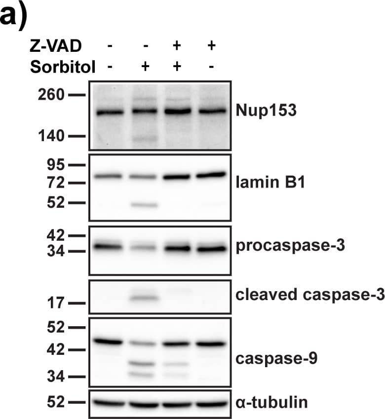
Detection of Human Caspase-9 by Western Blot Sorbitol-dependent TDP-43 relocalization is reversible and not dependent on Nup153 cleavage.(A) Western blot of nuclear pore complex protein Nup153, nuclear lamina component lamin B1, procaspases -3 and -9 and their activated forms caspase-3 and caspase-9 in HeLa cells treated with sorbitol after pre-treatment with DMSO control or caspase inhibitor Z-VAD-FMK. alpha -tubulin was used as a loading control. (B) Immunofluorescence staining of nuclear-cytoplasmic shuttling proteins TDP-43 (green) and HuR (red) in sorbitol-stressed HeLa cells after 0 min, 15 min or 60 min rescue in normal medium. Scale bar = 30 μm. (C) Quantification of cytoplasmic TDP-43 and HuR protein in sorbitol-treated HeLa cells after 0 min, 15 min or 60 min rescue (N = 3). Results represent mean percentage of cytoplasmic signal ± SEM;** p < 0.01, *** p < 0.001, **** p < 0.0001. Image collected and cropped by CiteAb from the following publication (https://pubmed.ncbi.nlm.nih.gov/28510586), licensed under a CC-BY license. Not internally tested by R&D Systems.
 View Larger
Detection of Human Human Caspase-9 Antibody by Western Blot Sorbitol-dependent TDP-43 relocalization is reversible and not dependent on Nup153 cleavage.(A) Western blot of nuclear pore complex protein Nup153, nuclear lamina component lamin B1, procaspases -3 and -9 and their activated forms caspase-3 and caspase-9 in HeLa cells treated with sorbitol after pre-treatment with DMSO control or caspase inhibitor Z-VAD-FMK. alpha -tubulin was used as a loading control. (B) Immunofluorescence staining of nuclear-cytoplasmic shuttling proteins TDP-43 (green) and HuR (red) in sorbitol-stressed HeLa cells after 0 min, 15 min or 60 min rescue in normal medium. Scale bar = 30 μm. (C) Quantification of cytoplasmic TDP-43 and HuR protein in sorbitol-treated HeLa cells after 0 min, 15 min or 60 min rescue (N = 3). Results represent mean percentage of cytoplasmic signal ± SEM;** p < 0.01, *** p < 0.001, **** p < 0.0001. Image collected and cropped by CiteAb from the following publication (https://pubmed.ncbi.nlm.nih.gov/28510586), licensed under a CC-BY license. Not internally tested by R&D Systems.
 View Larger
Detection of Human Caspase-9 by Western Blot Sorbitol-dependent TDP-43 relocalization is reversible and not dependent on Nup153 cleavage.(A) Western blot of nuclear pore complex protein Nup153, nuclear lamina component lamin B1, procaspases -3 and -9 and their activated forms caspase-3 and caspase-9 in HeLa cells treated with sorbitol after pre-treatment with DMSO control or caspase inhibitor Z-VAD-FMK. alpha -tubulin was used as a loading control. (B) Immunofluorescence staining of nuclear-cytoplasmic shuttling proteins TDP-43 (green) and HuR (red) in sorbitol-stressed HeLa cells after 0 min, 15 min or 60 min rescue in normal medium. Scale bar = 30 μm. (C) Quantification of cytoplasmic TDP-43 and HuR protein in sorbitol-treated HeLa cells after 0 min, 15 min or 60 min rescue (N = 3). Results represent mean percentage of cytoplasmic signal ± SEM;** p < 0.01, *** p < 0.001, **** p < 0.0001. Image collected and cropped by CiteAb from the following open publication (https://pubmed.ncbi.nlm.nih.gov/28510586), licensed under a CC-BY license. Not internally tested by R&D Systems.
Preparation and Storage
- 12 months from date of receipt, -20 to -70 °C as supplied.
- 1 month, 2 to 8 °C under sterile conditions after reconstitution.
- 6 months, -20 to -70 °C under sterile conditions after reconstitution.
Background: Caspase-9
Caspase-9 (Cysteine-aspartic acid protease 9/Casp-9; also APAF-3, Mch6 and ICE-LAP6) is a 35-37 kDa member of the peptidase C14A family of enzymes. Casp-9 is an initiator caspase that is part of the intrinsic apoptosis pathway. It is widely expressed and is particularly important during development. Human proCaspase-9 is a 47-48 kDa, 416 amino acid (aa) protein and it contains one CARD region (aa 1-92) and catalytic residues at His237 and Cys287. Following mitochondrial disruption, cytochrome c is released from mitochrondria. Cytochrome c acts on APAF-1, which induces procaspase-9 dimerization. The act of dimerization activates proCasp-9, leading to either the activation of Casp-3, or the autocleavage of proCasp-9, generating a 35 kDa subunit (aa 1-315) and a 12 kDa subunit. Activated Casp-3 will also act on proCasp-9, generating a 37 kDa subunit (aa 1-330) and a 10 kDa subunit (aa 331-416). These subunits associate to form an active heterotetramer. Casp-9 has an alternative start site at Met84 and a deletion of aa 140-289 that generates a dominant negative, 31 kDa isoform. Over aa 1-134, human Casp-9 shares 81% aa identity with mouse Casp-9.
Product Datasheets
Citations for Human Caspase-9 Antibody
R&D Systems personnel manually curate a database that contains references using R&D Systems products. The data collected includes not only links to publications in PubMed, but also provides information about sample types, species, and experimental conditions.
9
Citations: Showing 1 - 9
Filter your results:
Filter by:
-
Arachidin-1, a Prenylated Stilbenoid from Peanut, Enhances the Anticancer Effects of Paclitaxel in Triple-Negative Breast Cancer Cells
Authors: S Mohammadho, A Weaver, M Sudhakaran, LC Ho, T Le, AI Doseff, F Medina-Bol
Cancers, 2023-01-07;15(2):.
Species: Human
Sample Types: Cell Lysates
Applications: Western Blot -
Arachidin-1, a Prenylated Stilbenoid from Peanut, Induces Apoptosis in Triple-Negative Breast Cancer Cells
Authors: S Mohammadho, LC Ho, L Fang, J Xu, F Medina-Bol
International Journal of Molecular Sciences, 2022-01-20;23(3):.
Species: Human
Sample Types: Cell Lysates
Applications: Western Blot -
Opposing effects of polysulfides and thioredoxin on apoptosis through caspase persulfidation
Authors: I Braunstein, R Engelman, O Yitzhaki, T Ziv, E Galardon, M Benhar
J. Biol. Chem., 2020-02-10;0(0):.
Species: Human
Sample Types: Cell Lysates
Applications: Western Blot -
Truncation of the TAR DNA-binding protein 43 is not a prerequisite for cytoplasmic relocalization, and is suppressed by caspase inhibition and by introduction of the A90V sequence variant
Authors: HJ Wobst, L Delsing, NJ Brandon, SJ Moss
PLoS ONE, 2017-05-16;12(5):e0177181.
Species: Human
Sample Types: Cell Lysates
Applications: Western Blot -
Discovery of Sanggenon G as a natural cell-permeable small-molecular weight inhibitor of X-linked inhibitor of apoptosis protein (XIAP).
Authors: Seiter M, Salcher S, Rupp M, Hagenbuchner J, Kiechl-Kohlendorfer U, Mortier J, Wolber G, Rollinger J, Obexer P, Ausserlechner M
FEBS Open Bio, 2014-07-05;4(0):659-71.
Species: Human
Sample Types: Cell Lysates
Applications: Western Blot -
High-mobility group A1 protein inhibits p53-mediated intrinsic apoptosis by interacting with Bcl-2 at mitochondria
Authors: F Esposito, M Tornincasa, A Federico, G Chiappetta, G M Pierantoni, A Fusco
Cell Death & Disease
-
Intracellular K(+) inhibits apoptosis by suppressing the Apaf-1 apoptosome formation and subsequent downstream pathways but not cytochrome c release.
Authors: Karki P, Seong C, Kim JE, Hur K, Shin SY, Lee JS, Cho B, Park IS
Cell Death Differ., 2007-09-21;14(12):2068-75.
Species: Human
Sample Types: Cell Lysates
Applications: Western Blot -
Hsp70 inhibits heat-induced apoptosis upstream of mitochondria by preventing Bax translocation.
Authors: Stankiewicz A, Lachapelle G, Foo C, Radicioni S, Mosser D
J Biol Chem, 2005-09-19;280(46):38729-39.
Species: Human
Sample Types: Cell Lysates
Applications: Western Blot -
X-linked inhibitor of apoptosis (XIAP) confers human trophoblast cell resistance to Fas-mediated apoptosis.
Authors: Straszewski-Chavez S, Abrahams V, Funai E, Mor G
Mol Hum Reprod, 2004-01-01;10(1):33-41.
Species: Human
Sample Types: Cell Lysates
Applications: Western Blot
FAQs
No product specific FAQs exist for this product, however you may
View all Antibody FAQsReviews for Human Caspase-9 Antibody
Average Rating: 5 (Based on 1 Review)
Have you used Human Caspase-9 Antibody?
Submit a review and receive an Amazon gift card.
$25/€18/£15/$25CAN/¥75 Yuan/¥2500 Yen for a review with an image
$10/€7/£6/$10 CAD/¥70 Yuan/¥1110 Yen for a review without an image
Filter by:


