Human Caspase-3 Antibody Summary
Applications
Please Note: Optimal dilutions should be determined by each laboratory for each application. General Protocols are available in the Technical Information section on our website.
Scientific Data
 View Larger
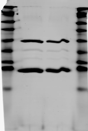
Detection of Human Precursor Caspase‑3 and p18 Subunit by Western Blot. Western blot shows lysates of Jurkat human acute T cell leukemia cell line untreated (-) or treated (+) with 1 µM staurosporine (STS) for 3 hours. PVDF membrane was probed with 1 µg/mL of Human Caspase-3 Monoclonal Antibody (Catalog # MAB707), followed by HRP-conjugated Anti-Mouse IgG Secondary Antibody (Catalog # HAF007). Specific bands were detected for precursor Caspase-3 and the p18 subunit at approximately 36 and 18 kDa (as indicated). This experiment was conducted under reducing conditions and using Immunoblot Buffer Group 4.
 View Larger
Western Blot Shows Human Caspase‑3 Specificity by Using Knockout Cell Line. Western blot shows lysates of HeLa human cervical epithelial carcinoma parental cell line and Caspase-3 knockout HeLa cell line (KO). PVDF membrane was probed with 2 µg/mL of Mouse Anti-Human Caspase-3 Monoclonal Antibody (Catalog # MAB707) followed by HRP-conjugated Anti-Mouse IgG Secondary Antibody (Catalog # HAF018). A specific band was detected for Caspase-3 at approximately 32 kDa (as indicated) in the parental HeLa cell line, but is not detectable in knockout HeLa cell line. GAPDH (Catalog # MAB5718) is shown as a loading control. This experiment was conducted under reducing conditions and using Immunoblot Buffer Group 1.
Preparation and Storage
- 12 months from date of receipt, -20 to -70 degreesC as supplied. 1 month, 2 to 8 degreesC under sterile conditions after reconstitution. 6 months, -20 to -70 degreesC under sterile conditions after reconstitution.
Background: Caspase-3
Caspase-3 (Cysteine-aspartic acid protease 3/Casp3; also Yama, apopain and CPP32) is a 29 kDa heterodimer that belongs to the peptidase C14A family of enzymes. It is widely expressed, and considered to be the major executioner caspase in the apoptotic cascade. Human procaspase-3 is a 32 kDa, 277 amino acid (aa) protein and is normally an inactive homodimer. Following cell stress/activation, procaspase-3 undergoes proteolysis to generate an N-terminal 148 aa p17/17 kDa subunit (aa 29-175), plus a 102 aa C-terminal p12/12 kDa subunit. These subunits noncovalently heterodimerize, and associate with another p17/p12 heterodimer to form an active enzyme. There is one potential variant that shows an alternative start site nine aa upstream of the standard start site coupled with a 21 aa substitution for aa 162-277. Over aa 29-175, human and mouse caspase-3 share 87% aa identity.
Product Datasheets
Citations for Human Caspase-3 Antibody
R&D Systems personnel manually curate a database that contains references using R&D Systems products. The data collected includes not only links to publications in PubMed, but also provides information about sample types, species, and experimental conditions.
8
Citations: Showing 1 - 8
Filter your results:
Filter by:
-
Interferon-? Overexpression in Adipose Tissue-Derived Stem Cells Induces HepG2 and Macrophage Cell Death in Liver Tumor Organoids via Induction of TNF-Related Apoptosis-Inducing Ligand Expression
Authors: Yoon, Y;Kim, CW;Kim, MY;Baik, SK;Jung, PY;Eom, YW;
International journal of molecular sciences
Species: Human
Sample Types: Organoids
Applications: Western Blot -
Alpha1-antitrypsin protects lung cancer cells from staurosporine-induced apoptosis: the role of bacterial lipopolysaccharide
Authors: N Schwarz, S Tumpara, S Wrenger, E Ercetin, J Hamacher, T Welte, S Janciauski
Sci Rep, 2020-06-12;10(1):9563.
Species: Human
Sample Types: Cell Culture Supernates
Applications: Western Blot -
Preeclampsia is associated with alterations in the p53-pathway in villous trophoblast.
Authors: Sharp A, Heazell A, Baczyk D, Dunk C, Lacey H, Jones C, Perkins J, Kingdom J, Baker P, Crocker I
PLoS ONE, 2014-01-30;9(1):e87621.
Species: Human
Sample Types: Tissue Homogenates
Applications: Western Blot -
PMA synergistically enhances apicularen A-induced cytotoxicity by disrupting microtubule networks in HeLa cells.
Authors: Seo K, Kim J, Park J, Song K, Yun E, Park J, Kweon G, Yoon W, Lim K, Hwang B
BMC Cancer, 2014-01-22;14(0):36.
Species: Human
Sample Types: Cell Lysates
Applications: Western Blot -
Interactions between important regulatory proteins and human alphaB crystallin.
Authors: Ghosh JG, Shenoy AK, Clark JI
Biochemistry, 2007-05-08;46(21):6308-17.
Species: Human
Sample Types: Recombinant Protein
Applications: ELISA-Based Protein Pin Array -
Developmental differences in the responses of IL-6 and IL-13 transgenic mice exposed to hyperoxia.
Authors: Choo-Wing R, Nedrelow JH, Homer RJ, Elias JA, Bhandari V
Am. J. Physiol. Lung Cell Mol. Physiol., 2007-03-30;293(1):L142-50.
Species: Mouse
Sample Types: Whole Tissue
Applications: IHC-P -
Proteolytically processed soluble tumor endothelial marker (TEM) 5 mediates endothelial cell survival during angiogenesis by linking integrin alpha(v)beta3 to glycosaminoglycans.
Authors: Vallon M, Essler M
J. Biol. Chem., 2006-09-17;281(45):34179-88.
Species: Human
Sample Types: Cell Lysates
Applications: Western Blot -
Inhibition of lymphotoxin-beta receptor-mediated cell death by survivin-DeltaEx3.
Authors: You RI, Chou YC
Cancer Res., 2006-03-15;66(6):3051-61.
Species: Human
Sample Types: Cell Lysates
Applications: Immunoprecipitation
FAQs
No product specific FAQs exist for this product, however you may
View all Antibody FAQsReviews for Human Caspase-3 Antibody
Average Rating: 5 (Based on 1 Review)
Have you used Human Caspase-3 Antibody?
Submit a review and receive an Amazon gift card.
$25/€18/£15/$25CAN/¥75 Yuan/¥2500 Yen for a review with an image
$10/€7/£6/$10 CAD/¥70 Yuan/¥1110 Yen for a review without an image
Filter by:


