Human Cadherin-13 Antibody Summary
Glu23-Ala692
Accession # P55290
Applications
Please Note: Optimal dilutions should be determined by each laboratory for each application. General Protocols are available in the Technical Information section on our website.
Scientific Data
 View Larger
Detection of Human Cadherin‑13 by Western Blot. Western blot shows lysates of human brain (cortex) tissue. PVDF Membrane was probed with 1 µg/mL of Goat Anti-Human Cadherin-13 Antigen Affinity-purified Polyclonal Antibody (Catalog # AF3264) followed by HRP-conjugated Anti-Goat IgG Secondary Antibody (Catalog # HAF019). A specific band was detected for Cadherin-13 at approximately 105 kDa (as indicated). This experiment was conducted under reducing conditions and using Immunoblot Buffer Group 8.
 View Larger
Cadherin‑13 in NCI-H460 Human Cell Line. Cadherin-13 was detected in immersion fixed NCI-H460 human large cell lung carcinoma cell line using 10 µg/mL Goat Anti-Human Cadherin-13 Antigen Affinity-purified Polyclonal Antibody (Catalog # AF3264) for 3 hours at room temperature. Cells were stained with the NorthernLights™ 557-conjugated Anti-Goat IgG Secondary Antibody (red; Catalog # NL001) and counter-stained with DAPI (blue). View our protocol for Fluorescent ICC Staining of Cells on Coverslips.
 View Larger
Cadherin‑13 in Human Skin. Cadherin-13 was detected in immersion fixed paraffin-embedded sections of human skin using Goat Anti-Human Cadherin-13 Antigen Affinity-purified Polyclonal Antibody (Catalog # AF3264) at 0.1 µg/mL overnight at 4 °C. Tissue was stained using the Anti-Goat HRP-DAB Cell & Tissue Staining Kit (brown; Catalog # CTS008) and counterstained with hematoxylin (blue). Specific staining was localized to plasma membranes of keratinocytes. View our protocol for Chromogenic IHC Staining of Paraffin-embedded Tissue Sections.
 View Larger
Detection of Human Cadherin‑13 by Simple WesternTM. Simple Western lane view shows lysates of human brain (motor cortex) tissue, loaded at 0.2 mg/mL. A specific band was detected for Cadherin-13 at approximately 114 kDa (as indicated) using 10 µg/mL of Goat Anti-Human Cadherin-13 Antigen Affinity-purified Polyclonal Antibody (Catalog # AF3264) followed by 1:50 dilution of HRP-conjugated Anti-Goat IgG Secondary Antibody (Catalog # HAF109). This experiment was conducted under reducing conditions and using the 12-230 kDa separation system.
 View Larger
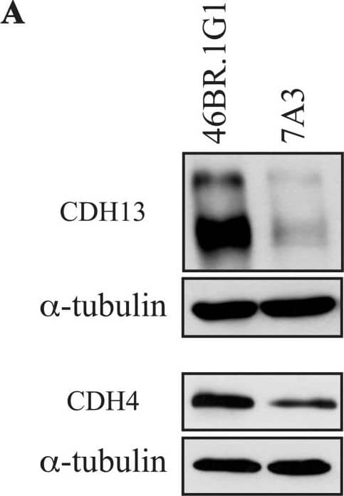
Detection of Human Cadherin-13 by Western Blot Differential expression of cadherin 13 and cadherin 4 proteins in 46BR.1G1 and 7A3 cells.(A) Cell lysates from 46BR.1G1 and 7A3 cells were analyzed by Western blotting with anti-cadherin 13, anti-cadherin 4, and anti-alpha -tubulin antibodies. (B) Quantification of the assay was performed by densitometric analysis with NIH ImageJ 1.43 program. Bars show mean ± SEM of three independent experiments. Image collected and cropped by CiteAb from the following publication (https://dx.plos.org/10.1371/journal.pone.0130561), licensed under a CC-BY license. Not internally tested by R&D Systems.
 View Larger
Detection of Human Cadherin-13 by Western Blot Differential expression of cadherin 13 and cadherin 4 proteins in 46BR.1G1 and 31W cells.Cell lysates from 46BR.1G1 and 31W cells were analyzed by Western blotting with antibodies against the indicated proteins. Image collected and cropped by CiteAb from the following publication (https://dx.plos.org/10.1371/journal.pone.0130561), licensed under a CC-BY license. Not internally tested by R&D Systems.
Preparation and Storage
- 12 months from date of receipt, -20 to -70 °C as supplied.
- 1 month, 2 to 8 °C under sterile conditions after reconstitution.
- 6 months, -20 to -70 °C under sterile conditions after reconstitution.
Background: Cadherin-13
Cadherin-13, also known as T-Cadherin and H-Cadherin, is a 105 kDa member of the cadherin family of transmembrane glycoproteins that mediate calcium-dependent intercellular adhesion (1). However, Cadherin-13 is an atypical member, lacking transmembrane and cytosolic domains and containing a GPI moiety that anchors Cadherin-13 to the plasma membrane (1‑2). Human Cadherin-13 is synthesized as a 713 amino acid (aa) precursor that contains a 22 aa signal sequence, a 116 aa propeptide, a 555 aa mature chain, and a second propeptide of 20 aa that is removed in the mature form to reveal the GPI anchor. The mature form contains five cadherin domains and eight potential sites for N-linked glycosylation. Mature human Cadherin-13 shares 96% aa identity with mature mouse Cadherin-13. Cadherin-13 is expressed in various tissues. It is highly expressed in the heart, and in the CNS, Cadherin-13 is expressed in the cerebral cortex, medulla, hippocampus, amygdala, thalamus, and substantia nigra (2). There are higher levels of Cadherin-13 in the adult brain than in developing brain (2). Cadherin‑13 is also expressed in skin in the basal layer of the epidermis, lung, liver, kidney, and blood vessels (2). The structural characteristics of Cadherin-13 predict that it is unlikely to function as a true adhesion molecule in vivo (2). It is suggested that it may act rather as a signaling receptor participating in recognition of the environment and regulation of cell motility, proliferation, and phenotype (2). Cellular expression levels of Cadherin‑13 in various tissues often correlate, negatively or positively, with the proliferative potential of the cells (2). Cadherin-13 may also act as a suppressor of tumor cell growth (2). This potential role for Cadherin‑13 was emphasized by localization of Cadherin-13 gene to chromosome 16q24, a region exhibiting loss of heterozygosity in many solid tumors (2). Allelic loss of chromosome bands 16q24.1-q24.2 and reduced expression of Cadherin‑13, as well as hypermethylation of the remaining allele have been detected in a considerable number of human cancers (2).
- Tanihara, H. et al. (1994) Cell Adhes. Commun. 2:15.
- Philippova, M. et al. (2009) Cell. Signal. 21:1035.
Product Datasheets
Citations for Human Cadherin-13 Antibody
R&D Systems personnel manually curate a database that contains references using R&D Systems products. The data collected includes not only links to publications in PubMed, but also provides information about sample types, species, and experimental conditions.
20
Citations: Showing 1 - 10
Filter your results:
Filter by:
-
Cadherin-13 Deficiency Increases Dorsal Raphe 5-HT Neuron Density and Prefrontal Cortex Innervation in the Mouse Brain
Authors: Andrea Forero, Olga Rivero, Sina Wäldchen, Hsing-Ping Ku, Dominik P. Kiser, Yvonne Gärtner et al.
Frontiers in Cellular Neuroscience
-
ER stress decreases exosome production through adiponectin/T-cadherin-dependent and -independent pathways
Authors: Keita Fukuoka, Ryohei Mineo, Shunbun Kita, Shiro Fukuda, Tomonori Okita, Emi Kawada-Horitani et al.
Journal of Biological Chemistry
-
Functional Properties of Rare Missense Variants of Human CDH13 Found in Adult Attention Deficit/Hyperactivity Disorder (ADHD) Patients
Authors: Thegna Mavroconstanti, Stefan Johansson, Ingeborg Winge, Per M. Knappskog, Jan Haavik
PLoS ONE
-
Chronic Replication Problems Impact Cell Morphology and Adhesion of DNA Ligase I Defective Cells
Authors: Cremaschi P, Oliverio M, Leva V et al.
PLoS ONE.
-
Pharmacological HIF-1 activation upregulates extracellular vesicle production synergistically with adiponectin through transcriptional induction and protein stabilization of T-cadherin
Authors: Fujii, K;Fujishima, Y;Kita, S;Kawada, K;Fukuoka, K;Sakaue, TA;Okita, T;Kawada-Horitani, E;Nagao, H;Fukuda, S;Maeda, N;Nishizawa, H;Shimomura, I;
Scientific reports
Species: Mouse
Sample Types: Cell Lysates
Applications: Western Blot -
Generation of induced pluripotent stem cell (iPSC) lines carrying a heterozygous (UKWMPi002-A-1) and null mutant knockout (UKWMPi002-A-2) of Cadherin 13 associated with neurodevelopmental disorders using CRISPR/Cas9
Authors: MR Vitale, JEM Zöller, C Jansch, A Janz, F Edenhofer, E Klopocki, D van den Ho, T Vanmierlo, O Rivero, N Nadif Kasr, GC Ziegler, KP Lesch
Stem Cell Research, 2021-01-11;51(0):102169.
Species: Human
Sample Types: Cell Lysates, Whole Cells
Applications: ICC, Western Blot -
Adiponectin Stimulates Exosome Release to Enhance Mesenchymal Stem-Cell-Driven Therapy of Heart Failure in Mice
Authors: Y Nakamura, S Kita, Y Tanaka, S Fukuda, Y Obata, T Okita, H Nishida, Y Takahashi, Y Kawachi, Y Tsugawa-Sh, Y Fujishima, H Nishizawa, Y Takakura, S Miyagawa, Y Sawa, N Maeda, I Shimomura
Mol. Ther., 2020-07-10;0(0):.
Species: Human
Sample Types: Cell Lysates
Applications: Western Blot -
Pioglitazone strengthen therapeutic effect of adipose-derived regenerative cells against ischemic cardiomyopathy through enhanced expression of adiponectin and modulation of macrophage phenotype
Authors: D Mori, S Miyagawa, R Matsuura, N Sougawa, S Fukushima, T Ueno, K Toda, T Kuratani, K Tomita, N Maeda, I Shimomura, Y Sawa
Cardiovasc Diabetol, 2019-03-22;18(1):39.
Species: Rat
Sample Types: Tissue Homogenates
Applications: Western Blot -
T-Cadherin Expression in Actinic Keratosis Transforming to Invasive Squamous Cell Carcinoma
Authors: Stanislaw A. Buechner, Therese J. Resink
Dermatopathology (Basel)
Species: Human
Sample Types: Whole Tissue
Applications: Immunohistochemistry -
Adiponectin/T-cadherin system enhances exosome biogenesis and decreases cellular ceramides by exosomal release
Authors: Y Obata, S Kita, Y Koyama, S Fukuda, H Takeda, M Takahashi, Y Fujishima, H Nagao, S Masuda, Y Tanaka, Y Nakamura, H Nishizawa, T Funahashi, B Ranscht, Y Izumi, T Bamba, E Fukusaki, R Hanayama, S Shimada, N Maeda, I Shimomura
JCI Insight, 2018-04-19;3(8):.
Species: Mouse
Sample Types: Cell Lysates, Whole Cells
Applications: ICC, Western Blot -
Actin cytoskeleton regulates functional anchorage-migration switch during T-cadherin-induced phenotype modulation of vascular smooth muscle cells
Authors: A Frismantie, E Kyriakakis, B Dasen, P Erne, TJ Resink, M Philippova
Cell Adh Migr, 2017-05-25;0(0):1-17.
Species: Human
Sample Types: Cell Lysates
Applications: Western Blot -
The unique prodomain of T-cadherin plays a key role in adiponectin binding with the essential extracellular cadherin repeats 1 and 2
Authors: S Fukuda, S Kita, Y Obata, Y Fujishima, H Nagao, S Masuda, Y Tanaka, H Nishizawa, T Funahashi, J Takagi, N Maeda, I Shimomura
J. Biol. Chem, 2017-03-21;0(0):.
Species: Mouse
Sample Types: Serum
Applications: Western Blot -
Vagal Nerve Stimulation Modifies Neuronal Activity and the Proteome of Excitatory Synapses of Amygdala/Piriform Cortex
Authors: Georgia M Alexander
J. Neurochem, 2017-02-03;0(0):.
Species: Rat
Sample Types: Protein
Applications: Western Blot -
T-Cadherin Expression in the Epidermis and Adnexal Structures of Normal Skin
Authors: Stanislaw Buechner
Dermatopathology (Basel), 2016-10-21;3(4):68-78.
Species: Human
Sample Types: Whole Tissue
Applications: IHC -
T-cadherin is essential for adiponectin-mediated revascularization.
Authors: Parker-Duffen, Jennifer, Nakamura, Kazuto, Silver, Marcy, Kikuchi, Ryosuke, Tigges, Ulrich, Yoshida, Sumiko, Denzel, Martin S, Ranscht, Barbara, Walsh, Kenneth
J Biol Chem, 2013-07-03;288(34):24886-97.
Species: Mouse
Sample Types: Whole Tissue
Applications: IHC-Fr, Immunoprecipitation, Western Blot -
Cross-talk between EGFR and T-cadherin: EGFR activation promotes T-cadherin localization to intercellular contacts.
Authors: Kyriakakis E, Maslova K, Frachet A, Ferri N, Contini A, Pfaff D, Erne P, Resink T, Philippova M
Cell Signal, 2013-02-11;25(5):1044-53.
Species: Human
Sample Types: Whole Cells
Applications: ICC, Western Blot -
Identification of proteins associating with glycosylphosphatidylinositol- anchored T-cadherin on the surface of vascular endothelial cells: role for Grp78/BiP in T-cadherin-dependent cell survival.
Authors: Philippova M, Ivanov D, Joshi MB, Kyriakakis E, Rupp K, Afonyushkin T, Bochkov V, Erne P, Resink TJ
Mol. Cell. Biol., 2008-04-14;28(12):4004-17.
Species: Human
Sample Types: Cell Lysates, Whole Cells
Applications: ICC, Western Blot -
Soluble T-cadherin promotes pancreatic beta -cell proliferation by upregulating Notch signaling
Authors: Tomonori Okita, Shunbun Kita, Shiro Fukuda, Keita Fukuoka, Emi Kawada-Horitani, Masahito Iioka et al.
iScience
-
Native adiponectin in serum binds to mammalian cells expressing T-cadherin, but not AdipoRs or calreticulin
Authors: Shunbun Kita, Shiro Fukuda, Norikazu Maeda, Iichiro Shimomura
eLife
-
T-Cadherin Expression in Actinic Keratosis Transforming to Invasive Squamous Cell Carcinoma
Authors: Stanislaw A. Buechner, Therese J. Resink
Dermatopathology (Basel)
FAQs
No product specific FAQs exist for this product, however you may
View all Antibody FAQsReviews for Human Cadherin-13 Antibody
There are currently no reviews for this product. Be the first to review Human Cadherin-13 Antibody and earn rewards!
Have you used Human Cadherin-13 Antibody?
Submit a review and receive an Amazon gift card.
$25/€18/£15/$25CAN/¥75 Yuan/¥2500 Yen for a review with an image
$10/€7/£6/$10 CAD/¥70 Yuan/¥1110 Yen for a review without an image
