Human Angiopoietin-1 Antibody Summary
Ser20-Phe498
Accession # Q15389
Applications
Please Note: Optimal dilutions should be determined by each laboratory for each application. General Protocols are available in the Technical Information section on our website.
Scientific Data
 View Larger
Angiopoietin‑1 in Human Prostate Cancer Tissue. Angiopoietin-1 was detected in immersion fixed paraffin-embedded sections of human prostate cancer tissue using Mouse Anti-Human Angiopoietin-1 Monoclonal Antibody (Catalog # MAB923) at 15 µg/mL overnight at 4 °C. Tissue was stained using the Anti-Mouse HRP-DAB Cell & Tissue Staining Kit (brown; Catalog # CTS002) and counterstained with hematoxylin (blue). Specific staining was localized to cytoplasm in cancer cells. View our protocol for Chromogenic IHC Staining of Paraffin-embedded Tissue Sections.
 View Larger
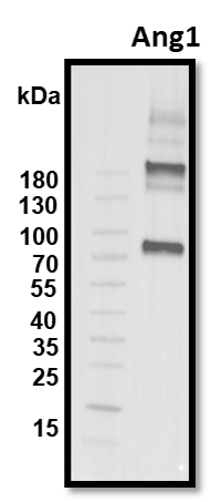
Detection of Mouse Angiopoietin-1 by Western Blot Pericyte-derived Angpt1 controls alveologenesis. a RT-qPCR analysis of Angpt1 and Tie2/Tek expression in freshly sorted lung GFP+, CD31+ or EpCAM+ cells from P7 Pdgfrb(BAC)-CreERT2 R26-mT/mG mice. Data represents mean ± s.e.m. (n = 4 mice). b High magnification images of P10 Angpt1GFP lungs stained for GFP (green), PDGFR beta (red), and PDGFR alpha (blue). Arrows indicate GFP and PDGFR beta double positive pericytes. Scale bar, 15 µm. c RT-qPCR analysis of Angpt1 expression in freshly sorted PDGFR beta + cells from P7 Yap1,Wwtr1iPCKO and control lungs. Data represents mean ± s.e.m. (n = 4 mice, two-tailed unpaired t-test). dAngpt1 expression in cultured Verteporfin (VP)-treated (48 h) and control pericytes. Data represents mean ± s.e.m. (n = 4, Welch’s t-test). e Expression of the indicated transcripts in freshly sorted CD31+ cells from P7 Yap1,Wwtr1iPCKO and control lungs. Data represents mean ± s.e.m. (n = 4 mice, NS not significant, two-tailed unpaired t-test). f–h Western blot analysis of Angpt1 protein (f; n = 2 controls and 4 mutant mice) and of total and phospho-Tie2 (pTie2) in P12 Yap1,Wwtr1iPCKO and control total lung lysates (g, n = 3 controls and 5 mutants). Molecular weight marker (kDa) is indicated. Relative quantification of signals is shown in h. Two-tailed unpaired t-test. i Scheme showing the time points of tamoxifen administration and analysis for Angpt1iPCKO mice. j, k 3D reconstruction confocal images of P12 Angpt1iPCKO and littermate control lungs stained for AQP5 (green), PDGFR beta (red), and PECAM1 (blue). Panels in k show higher magnification of PECAM1 staining. Scale bar, 50 µm (j) and 30 µm (k). l Quantitation of airspace volume in P12 Angpt1iPCKO and littermate control lung sections with 3D reconstruction surface images. Data represents mean ± s.e.m. (n = 4 mice; p < 0.0001, two-tailed unpaired t-test). m 3D reconstruction confocal images of P12 Angpt1iPCKO and littermate control lungs stained for alpha SMA (red) and tropoelastin (blue). Scale bar, 50 µm. n Quantitation of staining intensity for alpha SMA or tropoelastin shown in m. Intensity was normalized to the size of the parenchymal region. Data represents mean ± s.e.m. (n = 4 mice; NS not significant, two-tailed unpaired t-test). o Schematic summary of findings. Pulmonary pericytes regulate ECs and alveolar epithelial cells via angiocrine factors such as angiopoietin-1 and HGF Image collected and cropped by CiteAb from the following publication (https://pubmed.ncbi.nlm.nih.gov/29934496), licensed under a CC-BY license. Not internally tested by R&D Systems.
 View Larger
Detection of Porcine Angiopoietin-1 by Immunohistochemistry The Angpt2/Angpt1 balance is disturbed in atherogenic DM pigs.A. Representative illustrations of kidney sections stained with Angpt1 (upper panels; arrow: glomerulus; arrowhead: peritubular area) or Angpt2 (lower panels; arrow: glomerulus; arrowhead: tubular staining) in Controls, ATH, and DM+ATH pigs. B. Immunofluorescent double staining of representative kidney sections for desmin (green)/Angpt1 (red;left panel) and vWF (green/Angpt2 (red; middle/right panel). Insets: double positivity for vWF/Angpt2 staining in yellow. C: Quantitative analysis of renal expression of Angpt1, Angpt2 and Angpt2/Angpt1 ratio. D. Relative mRNA expression of Angpt1 and Angpt2. Data are shown as mean ± SEM. *P<0.05 compared to Controls or ATH pigs. Original magnification of A and B: x400. Image collected and cropped by CiteAb from the following publication (https://dx.plos.org/10.1371/journal.pone.0121555), licensed under a CC-BY license. Not internally tested by R&D Systems.
Preparation and Storage
- 12 months from date of receipt, -20 to -70 °C as supplied.
- 1 month, 2 to 8 °C under sterile conditions after reconstitution.
- 6 months, -20 to -70 °C under sterile conditions after reconstitution.
Background: Angiopoietin-1
Angiopoietin-1 (Ang-1) and Angiopoietin-2 (Ang-2) are two closely related secreted ligands which bind with similar affinity to Tie-2, a receptor tyrosine kinase with immunoglobulin and epidermal growth factor homology domains expressed primarily on endothelial cells and early hematopoietic cells. Tie-2 and angiopoietins have been shown to play critical roles in embryogenic angiogenesis and in maintaining the integrity of the adult vasculature (1). Ang-1 cDNA encodes a 498 amino acid (aa) precursor protein that contains a coiled-coiled domain near the amino-terminus and a fibrinogen-like domain at the C-terminus. Human Ang-1 shares approximately 97% and 60% aa sequence identity with mouse Ang‑1 and human Ang‑2, respectively (1, 2). Ang-1 activates Tie-2 signaling on endothelial cells to promote chemotaxis, cell survival, cell sprouting, vessel growth and stabilization (1, 3, 4). Ang-2 has alternatively been reported to be an antagonist for Ang-1-induced Tie-2 signaling as well as an agonist for Tie-2 signaling, depending on the cell context (5).
- Jones, N. et al. (2001) Nat. Rev. Mol. Cell Biol. 2:257.
- Davis, S. et al. (1996) Cell 87:1161.
- Witzenbichler, B. et al. (1998) J. Biol. Chem. 273:18514.
- Papapetropoulos, A. et al. (1999) Lab. Inest. 79:213.
- Teichert-Kuliszewska, K. et al. (2001) Cardiovasc. Res. 49:659.
Product Datasheets
Citations for Human Angiopoietin-1 Antibody
R&D Systems personnel manually curate a database that contains references using R&D Systems products. The data collected includes not only links to publications in PubMed, but also provides information about sample types, species, and experimental conditions.
10
Citations: Showing 1 - 10
Filter your results:
Filter by:
-
Non-neutralizing antibodies increase endogenous circulating Ang1 levels
Authors: Zheng C, Toth J, Bigwarfe T et al.
MAbs
-
Investigating the paracrine and juxtacrine abilities of adipose-derived stromal cells in angiogenesis triple cell co-cultures
Authors: Harary Søndergaard, R;Drozd Højgaard, L;Haack-Sørensen, M;Hoeeg, C;Mønsted Johansen, E;Follin, B;Kastrup, J;Ekblond, A;Juhl, M;
Stem cell research
Species: Human
Sample Types: Whole Cells
Applications: Neutralization -
Pulmonary pericytes regulate lung morphogenesis
Authors: K Kato, R Diéguez-Hu, DY Park, SP Hong, S Kato-Azuma, S Adams, M Stehling, B Trappmann, JL Wrana, GY Koh, RH Adams
Nat Commun, 2018-06-22;9(1):2448.
Species: Mouse
Sample Types: Cell Lysates
Applications: Western Blot -
Fibulin-5 Regulates Angiopoietin-1/Tie-2 Receptor Signaling in Endothelial Cells
Authors: Wilson Chan
PLoS ONE, 2016-06-15;11(6):e0156994.
Species: Human
Sample Types: Protein
Applications: ELISA Development -
Anti-tumoral, anti-angiogenic and anti-metastatic efficacy of a tetravalent bispecific antibody (TAvi6) targeting VEGF-A and angiopoietin-2
Authors: Werner Scheuer, Markus Thomas, Petra Hanke, Johannes Sam, Franz Osl, Diana Weininger et al.
mAbs
-
Early systemic microvascular damage in pigs with atherogenic diabetes mellitus coincides with renal angiopoietin dysbalance.
Authors: Khairoun M, van den Heuvel M, van den Berg B, Sorop O, de Boer R, van Ditzhuijzen N, Bajema I, Baelde H, Zandbergen M, Duncker D, Rabelink T, Reinders M, van der Giessen W, Rotmans J
PLoS ONE, 2015-04-24;10(4):e0121555.
Species: Porcine
Sample Types: Whole Tissue
Applications: IHC-Fr -
High bone marrow angiopoietin-1 expression is an independent poor prognostic factor for survival in patients with myelodysplastic syndromes.
Authors: Cheng CL, Hou HA, Jhuang JY, Lin CW, Chen CY, Tang JL, Chou WC, Tseng MH, Yao M, Huang SY, Ko BS, Hsu SC, Wu SJ, Tsay W, Chen YC, Tien HF
Br. J. Cancer, 2011-08-30;105(7):975-82.
Species: Human
Sample Types: Whole Tissue
Applications: IHC -
Integrin binding angiopoietin-1 monomers reduce cardiac hypertrophy.
Authors: Dallabrida SM, Ismail NS, Pravda EA, Parodi EM, Dickie R, Durand EM, Lai J, Cassiola F, Rogers RA, Rupnick MA
FASEB J., 2008-05-23;22(8):3010-23.
Species: Mouse, Rat
Sample Types: Cell Culture Supernates
Applications: Immunoprecipitation -
Regulation of angiopoietin expression by bacterial lipopolysaccharide.
Authors: Mofarrahi M, Nouh T, Qureshi S, Guillot L, Mayaki D, Hussain SN
Am. J. Physiol. Lung Cell Mol. Physiol., 2008-02-29;294(5):L955-63.
Species: Mouse
Sample Types: Tissue Homogenates
Applications: Western Blot -
Angiopoietin 1 directly induces destruction of the rheumatoid joint by cooperative, but independent, signaling via ERK/MAPK and phosphatidylinositol 3-kinase/Akt.
Authors: Hashiramoto A, Sakai C, Yoshida K, Tsumiyama K, Miura Y, Shiozawa K, Nose M, Komai K, Shiozawa S
Arthritis Rheum., 2007-07-01;56(7):2170-9.
Species: Human
Sample Types: Whole Cells, Whole Tissue
Applications: ICC, IHC
FAQs
No product specific FAQs exist for this product, however you may
View all Antibody FAQsReviews for Human Angiopoietin-1 Antibody
Average Rating: 4.5 (Based on 2 Reviews)
Have you used Human Angiopoietin-1 Antibody?
Submit a review and receive an Amazon gift card.
$25/€18/£15/$25CAN/¥75 Yuan/¥2500 Yen for a review with an image
$10/€7/£6/$10 CAD/¥70 Yuan/¥1110 Yen for a review without an image
Filter by:


