Hoechst 33342
Chemical Name: 2'-(4-Ethoxyphenyl)-5-(4-methyl-1-piperazinyl)-2,5'-bi-1H-benzimidazole trihydrochloride
Purity: ≥98%
Biological Activity
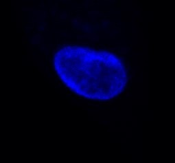
Key information: Hoechst 33342 is a blue-fluorescent dye for DNA staining. Cell permeable. Suitable for fixed and live-cell staining. Commonly used as a counterstain in fluorescence microscopy.Used for: nuclear counterstain, DNA visualization, cell cycle studies, apoptosis analysis.
Application: flow cytometry, confocal microscopy.
Properties and Photophysical Data: Hoechst 33342 binds to the AT-rich regions of the minor grove in DNA which renders it specific for nuclear chromatin. Excitation and emission maxima (λ) are 350 nm and 461 nm, respectively.
Scientific Data
 View Larger
Hoechst 33342 | CAS No. 875756-97-1 Fluorescent dye for staining DNA
Optical Data for Hoechst 33342
Plan Your Experiments
Use our spectra viewer to interactively plan your experiments, assessing multiplexing options. View the excitation and emission spectra for our fluorescent dye range and other commonly used dyes.
Spectral ViewerTechnical Data
The technical data provided above is for guidance only.
For batch specific data refer to the Certificate of Analysis.
Tocris products are intended for laboratory research use only, unless stated otherwise.
Background References
-
Assignment of DNA binding sites for 4',6-diamidine-2-phenylindole and bisbenzimide (Hoechst 33258). A comparative footprinting study.
Portugal and Waring
Biochim.Biophys.Acta., 1988;949:158 -
Simultaneous quantitation of Hoechst 33342 and immunofluorescence on viable cells using a fluorescence activated cell sorter.
Loken
Cytometry, 1980;1:136
Product Datasheets
Citations for Hoechst 33342
The citations listed below are publications that use Tocris products. Selected citations for Hoechst 33342 include:
12 Citations: Showing 1 - 10
-
A Simple and Affordable Method to Create Nonsense Mutation Clones of p53 for Studying the Premature Termination Codon Readthrough Activity of PTC124.
Authors: Ke-Fang Et al.
Biomedicines 2023;11
-
Methods for mitochondrial health assessment by High Content Imaging System.
Authors: Chayanon Et al.
MethodsX 2022;9:101685
-
A Multiscale Study of Phosphorylcholine Driven Cellular Phenotypic Targeting.
Authors: Diana Et al.
ACS Cent Sci 2022;8:891-904
-
Receptor Interactome Discovery with RDIMIS, a Membrane Protein Interaction Screen Using Recombinant Extracellular Vesicles.
Authors: Mike Et al.
Curr Protoc 2022;2:e483
-
Involvement of P2Y12 receptors in a nitroglycerin-induced model of migraine in male mice.
Authors: Mária Et al.
Br J Pharmacol 2021;178:4626-4645
-
Mechanical Counterbalance of Kinesin and Dynein Motors in a Microtubular Network Regulates Cell Mechanics, 3D Architecture, and Mechanosensing.
Authors: Jian Et al.
ACS Nano 2021;15:17528-17548
-
Human Breast Extracellular Matrix Microstructures and Protein Hydrogel 3D Cultures of Mammary Epithelial Cells.
Authors: Steve R Et al.
Cancers (Basel) 2021;13
-
Engineering Elastic Nano- and Micro-Patterns and Textures for Directed Cell Motility.
Authors: Paolo P Et al.
STAR Protoc 2020;1
-
SIGMAR1/Sigma-1 receptor ablation impairs autophagosome clearance.
Authors: Yang Et al.
Autophagy 2019;:1
-
GORASP2/GRASP55 collaborates with the PtdIns3K UVRAG complex to facilitate autophagosome-lysosome fusion.
Authors: Jie Et al.
Autophagy 2019;15:1787-1800
-
Subcutaneous Inoculation of 3D Pancreatic Cancer Spheroids Results in Development of Reproducible Stroma-Rich Tumors.
Authors: Durymanov Et al.
Transl Oncol 2019;12:180
-
Microtubule-Actomyosin Mechanical Cooperation during Contact Guidance Sensing.
Authors: Tabdanov Et al.
Cell Rep 2018;25:328
FAQs
No product specific FAQs exist for this product, however you may
View all Small Molecule FAQsReviews for Hoechst 33342
Average Rating: 5 (Based on 3 Reviews)
Have you used Hoechst 33342?
Submit a review and receive an Amazon gift card.
$25/€18/£15/$25CAN/¥75 Yuan/¥2500 Yen for a review with an image
$10/€7/£6/$10 CAD/¥70 Yuan/¥1110 Yen for a review without an image
Filter by:
Hoechst was used for nuclear staining. Add stain at 1 ug/mL and incubate cells at room temperature for 10 minutes.
I used this dye to stain the nucleus of live cells. After making the stock solution, diluted the dye 1:1000 ratio in the culture media/PBS and added to cells and incubated for 5 mins.


